Copyright
©The Author(s) 2022.
World J Gastrointest Oncol. Aug 15, 2022; 14(8): 1552-1561
Published online Aug 15, 2022. doi: 10.4251/wjgo.v14.i8.1552
Published online Aug 15, 2022. doi: 10.4251/wjgo.v14.i8.1552
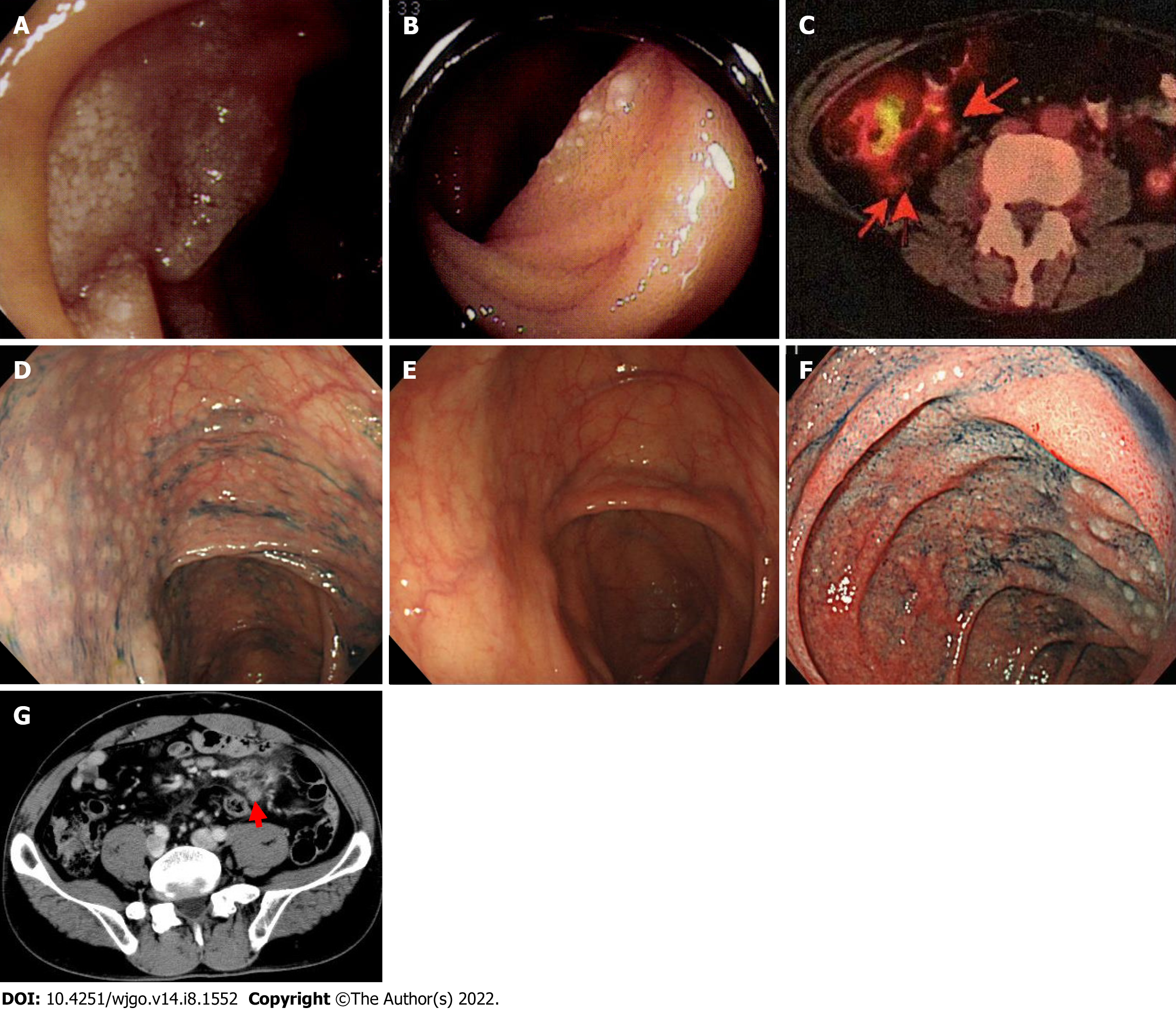
Figure 1 Videography findings of cases 1-3.
A and B: Endoscopic findings of case 1, lesions at (A) the descending portion of the duodenum and (B) the jejunum; C-E: Positron emission tomography findings of case 2; the arrow indicates a mesenteric nodal lesion in the ileocecal region (C); colonoscopy findings showed multiple lymphomatous polyposis-like lesions in the ascending colon (D); 1 year later, the lesion spontaneously disappeared (E); F and G: Esophagogastroduodenoscopy findings of case 3; lymphoma lesions were revealed in the descending portion of the duodenum (F); abdominal computed tomography findings. Mesenteric lymph nodes were swollen (arrowhead) (G).
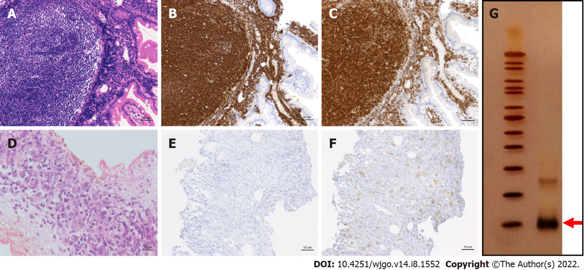
Figure 2 Pathological findings of case 5.
The upper row shows the histology at the time of onset; the lower row shows images obtained 21 years later and at the final stage of treatment; A and D: Hematoxylin and eosin staining; B and E: CD20 staining; C and F: BCL-2 staining; G: The PCR-single strand conformation polymorphism method. The arrow indicates bands that represent B-cell clones.
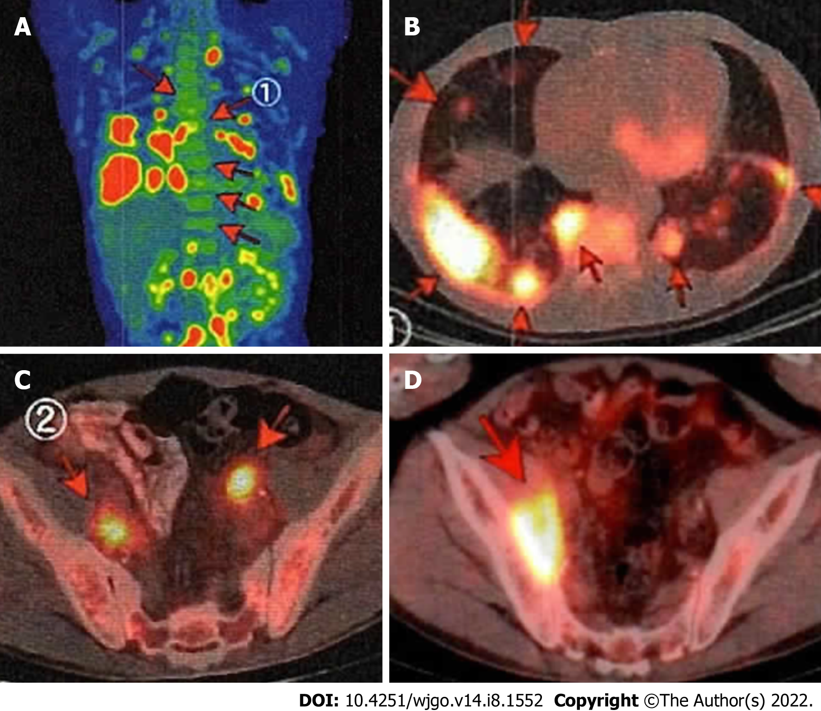
Figure 3 Positron emission tomography-computed tomography imaging of case 5.
Arrows point to lymphoma lesions at recurrence; A: Longitudinal image; B: Chest; C: Pelvic cavity; D: After treatment with various anticancer drugs, the nodal lesions in the pelvic cavity progressed further.
- Citation: Saito M, Mori A, Tsukamoto S, Ishio T, Yokoyama E, Izumiyama K, Morioka M, Kondo T, Sugino H. Duodenal-type follicular lymphoma more than 10 years after treatment intervention: A retrospective single-center analysis. World J Gastrointest Oncol 2022; 14(8): 1552-1561
- URL: https://www.wjgnet.com/1948-5204/full/v14/i8/1552.htm
- DOI: https://dx.doi.org/10.4251/wjgo.v14.i8.1552


