Copyright
©The Author(s) 2021.
World J Gastrointest Oncol. Aug 15, 2021; 13(8): 822-834
Published online Aug 15, 2021. doi: 10.4251/wjgo.v13.i8.822
Published online Aug 15, 2021. doi: 10.4251/wjgo.v13.i8.822
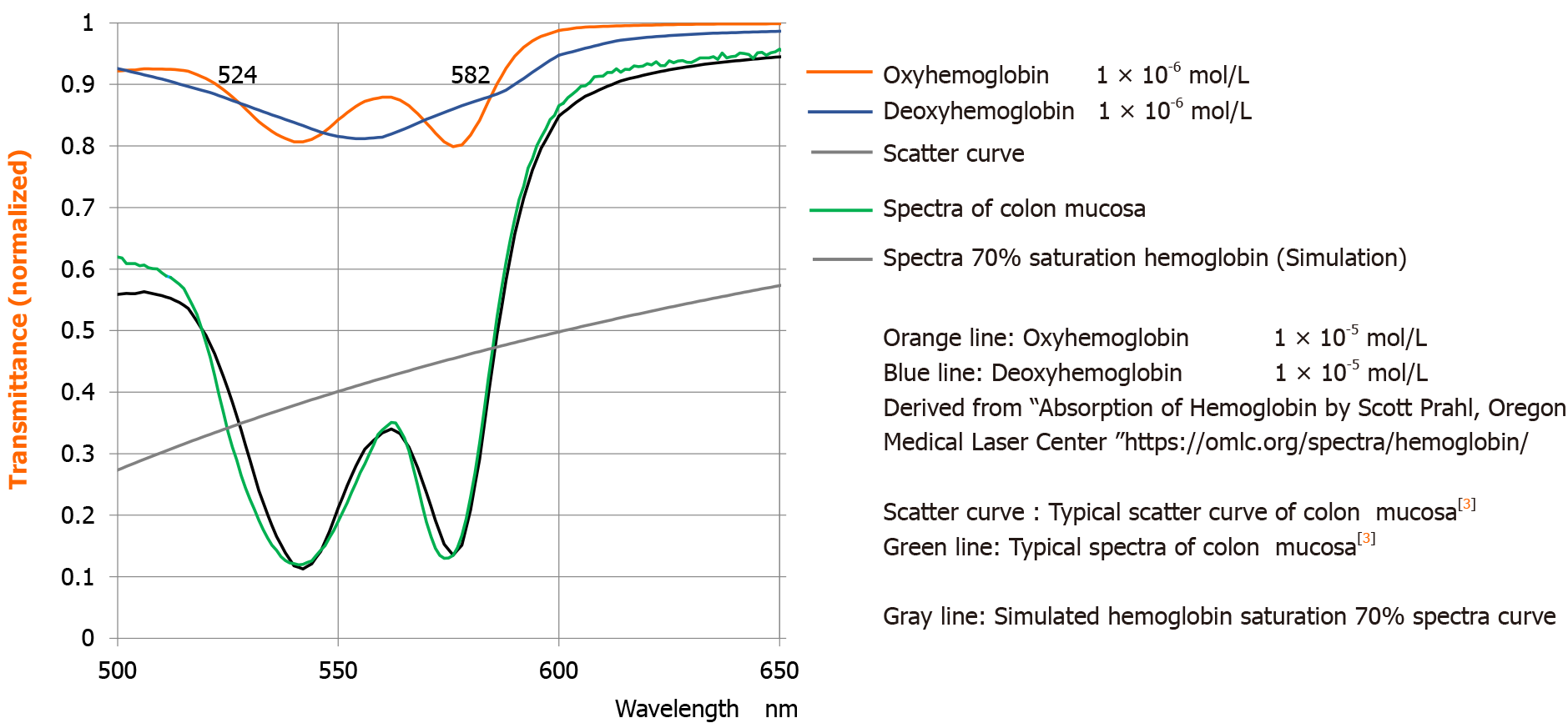
Figure 1
Spectra components oxyHb and deoxyHb and scatter.
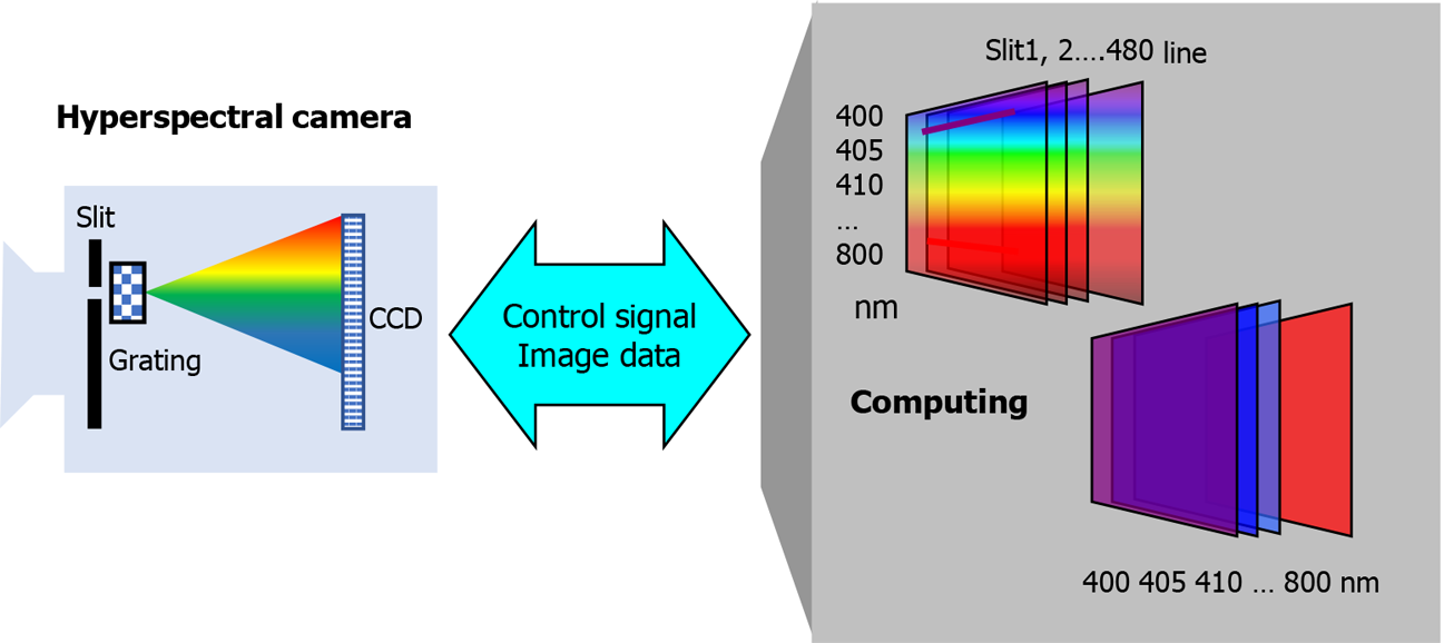
Figure 2
Scheme of hyperspectral camera.
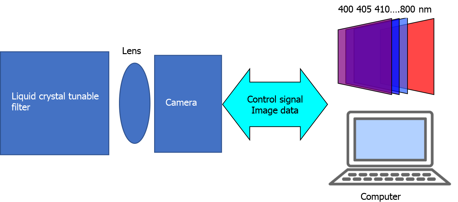
Figure 3
Scheme of liquid crystal tunable filter and digital camera combination.
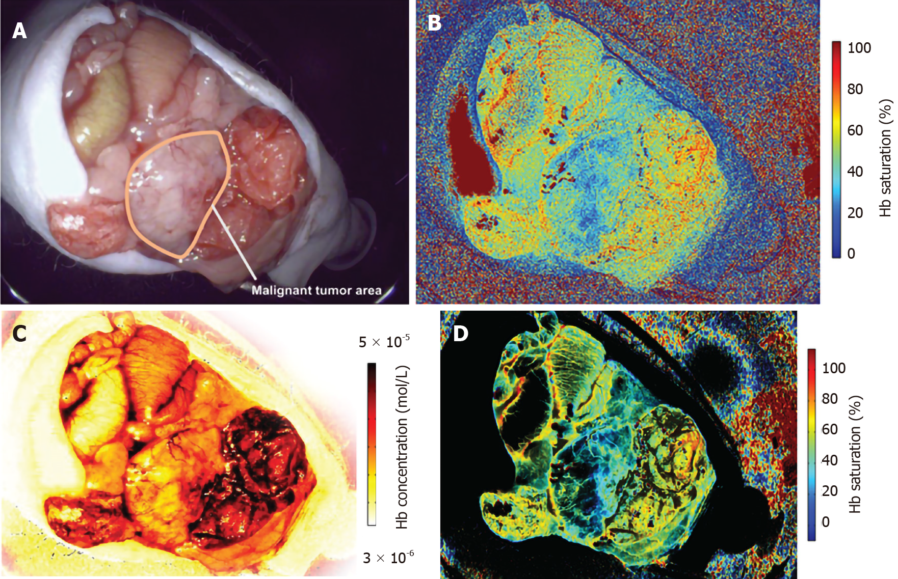
Figure 4 The system enables to indicate hemoglobin saturation of the mucosa with a practical frame rate for normal examination.
An area with relatively high Hb concentration area is enhanced by an Hb concentration mask. The higher Hb concentration and lower Hb saturation areas that are influenced by the tumor are observed as a blue area. A: Experimental cancer model. Red–green–blue video endoscope image of the mouse viscera. The tumor area is outlined by a white line; B: Hb saturation map of the tumor generated by the endoscope system; C: Hb concentration map of the tumor generated by the endoscope system; D: Hb concentration-masked Hb saturation map.
- Citation: Chiba T, Murata M, Kawano T, Hashizume M, Akahoshi T. Reflectance spectra analysis for mucous assessment. World J Gastrointest Oncol 2021; 13(8): 822-834
- URL: https://www.wjgnet.com/1948-5204/full/v13/i8/822.htm
- DOI: https://dx.doi.org/10.4251/wjgo.v13.i8.822


