Copyright
©The Author(s) 2020.
World J Gastrointest Oncol. Jul 15, 2020; 12(7): 705-718
Published online Jul 15, 2020. doi: 10.4251/wjgo.v12.i7.705
Published online Jul 15, 2020. doi: 10.4251/wjgo.v12.i7.705
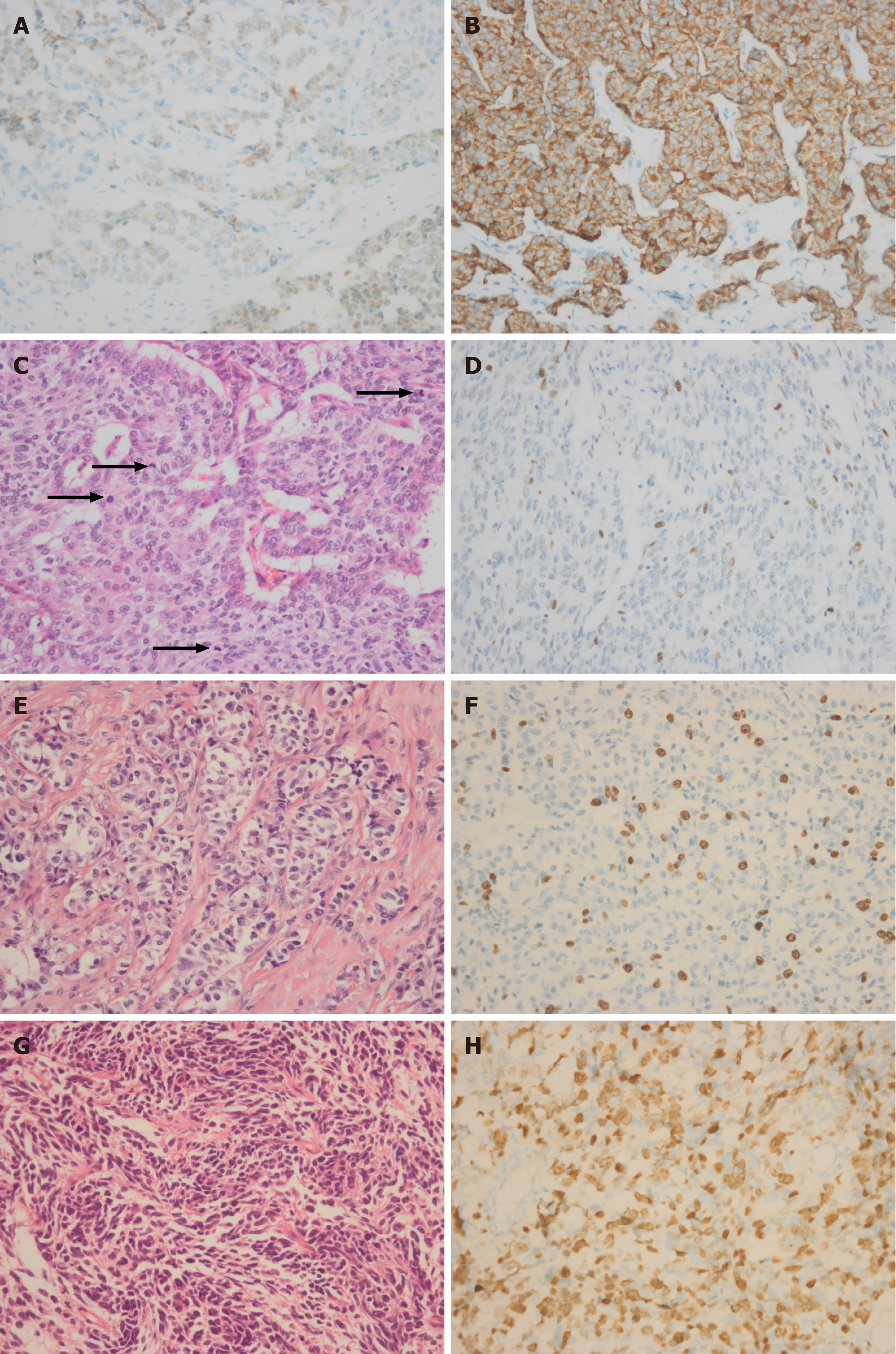
Figure 1 Immunohistochemical staining (400 ×).
A and B: Immunohistochemical positivity for chromogranin A (A) or synaptophysin (B) is considered to be a marker for neuroendocrine neoplasms. Mitotic figures rating [G3 26/10 high-power field (HPF)] is higher than the Ki67 index rating (G2, 16%); C and D: Four mitoses in one HPF is shown by black arrows (C) and related Ki67 immunohistochemical staining is shown in D; E and F: Hemtoxylin and eosin (H&E) staining of well-differentiated pancreatic neuroendocrine tumor G3 (E) and related Ki67 immunohistochemical staining (F); G and H: H&E staining of poorly differentiated pancreatic neuroendocrine carcinoma (G) and related Ki-67 immunohistochemical staining (H).
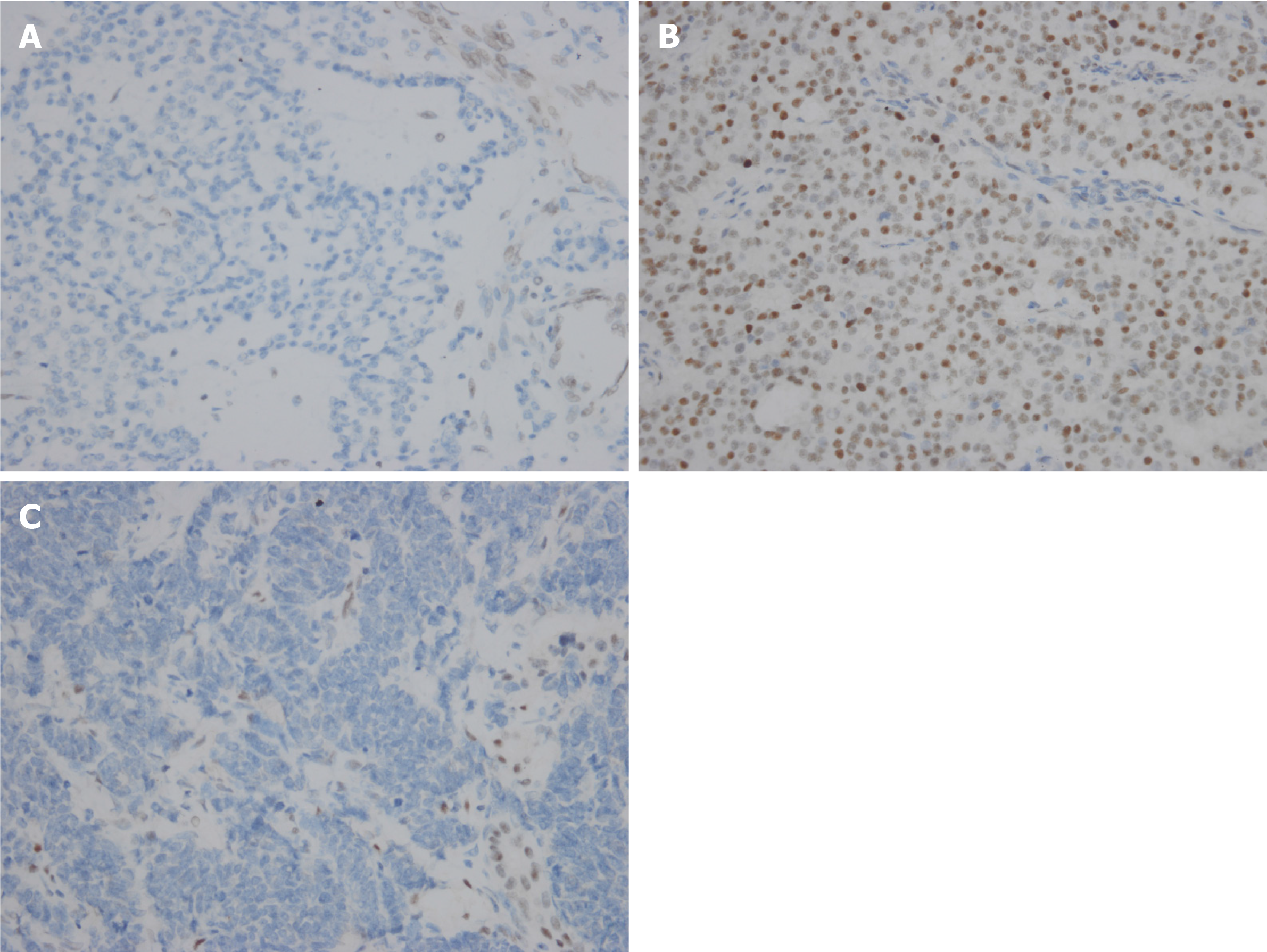
Figure 2 Immunohistochemical staining (400 ×).
A: Loss of nuclear expression for ATRX in pancreatic neuroendocrine tumor G3; B and C: Aberrant expression for p53 (B) and loss of expression of Rb1 (C) in pancreatic neuroendocrine carcinoma.
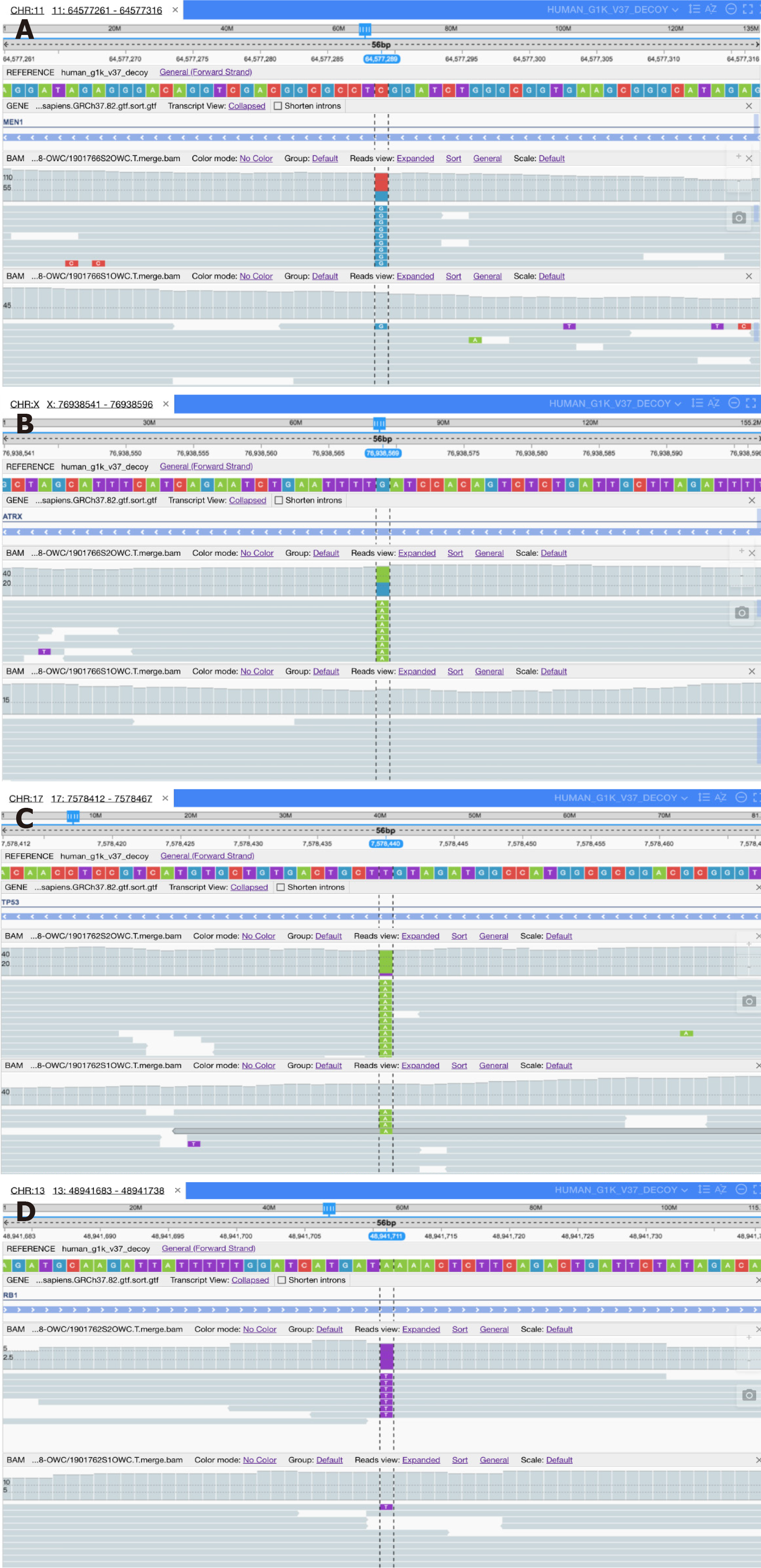
Figure 3 High-throughput sequencing.
A: Base C (red) mutates to G (blue) on the Men1 gene of chromosome 11 in pancreatic neuroendocrine tumor G3 (pNET G3); B: Base G (blue) mutates to A (green) on the ARTX gene of chromosome X in pNET G3; C: Base T (purple) mutates to A (green) on the Tp53 gene of chromosome 17 in pancreatic neuroendocrine carcinoma (pNEC); D: Base A (green) mutates to T (purple) on the Rb1 gene of chromosome 13 in pNEC.
- Citation: Zhang MY, He D, Zhang S. Pancreatic neuroendocrine tumors G3 and pancreatic neuroendocrine carcinomas: Differences in basic biology and treatment. World J Gastrointest Oncol 2020; 12(7): 705-718
- URL: https://www.wjgnet.com/1948-5204/full/v12/i7/705.htm
- DOI: https://dx.doi.org/10.4251/wjgo.v12.i7.705


