Copyright
©The Author(s) 2020.
World J Gastrointest Oncol. Mar 15, 2020; 12(3): 267-275
Published online Mar 15, 2020. doi: 10.4251/wjgo.v12.i3.267
Published online Mar 15, 2020. doi: 10.4251/wjgo.v12.i3.267
Figure 1 Overview of extracellular matrix preparation.
A: Normal human colon tissue; B: Human colorectal cancer tissue; C: Normal human colon tissue or human colorectal cancer tissue were decellularized and shown in a glass culture dish.
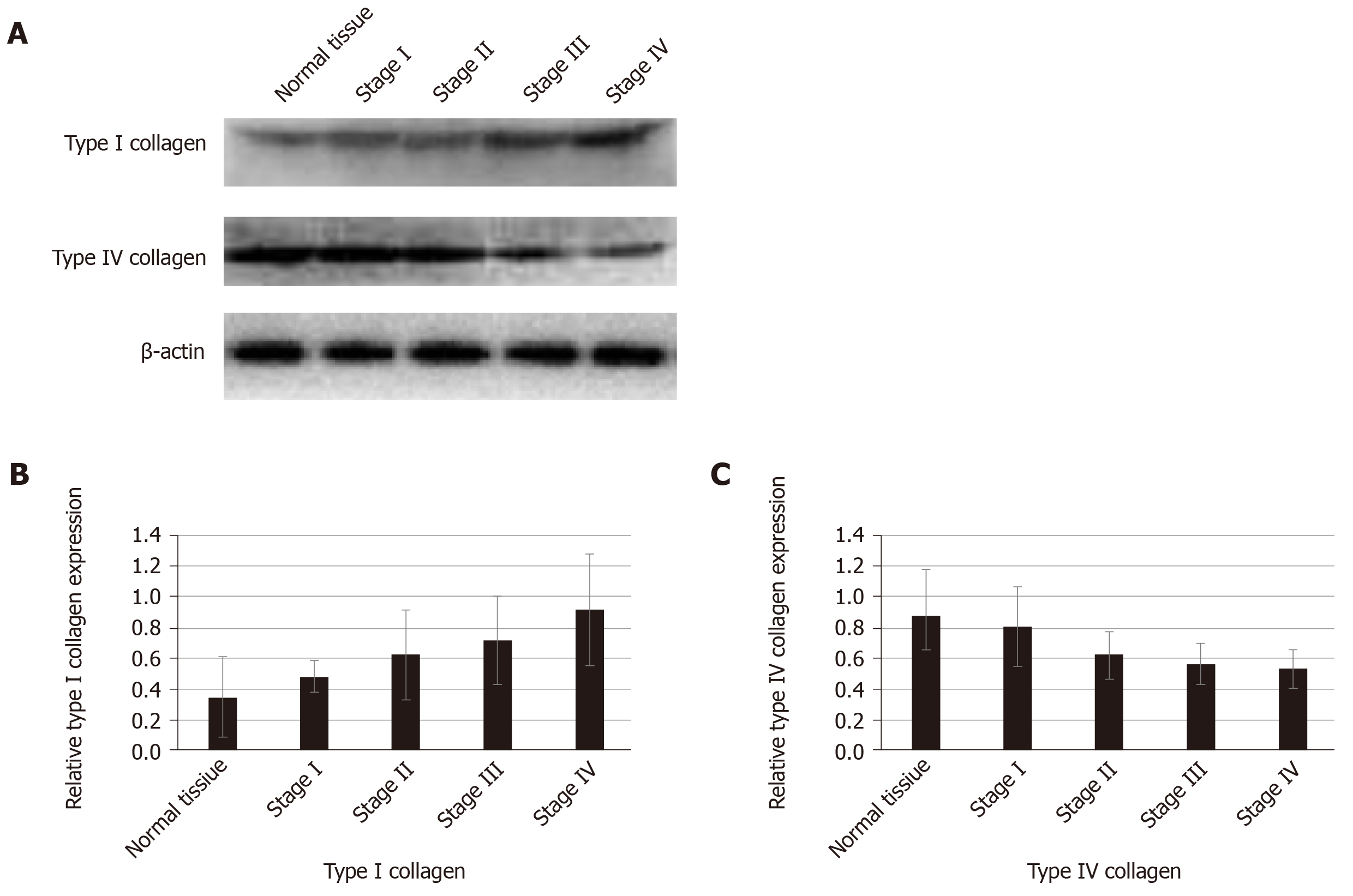
Figure 2 Expression of type I collagen and type IV collagen in normal tissue and colorectal cancer.
A: Western blot showing the expression of type I collagen and type IV collagen; B: The expression of type I collagen in the extracellular matrix (ECM) of stages III and IV colorectal cancer was highest. In the ECM of stages I and II colorectal cancer, the expression was relatively low. The expression of type I collagen was positively associated with the stage of colorectal cancer (r = 0.706, P < 0.01); C: The expression of type IV collagen was negatively correlated with the stage of colorectal cancer (r = - 0.796, P < 0.01).
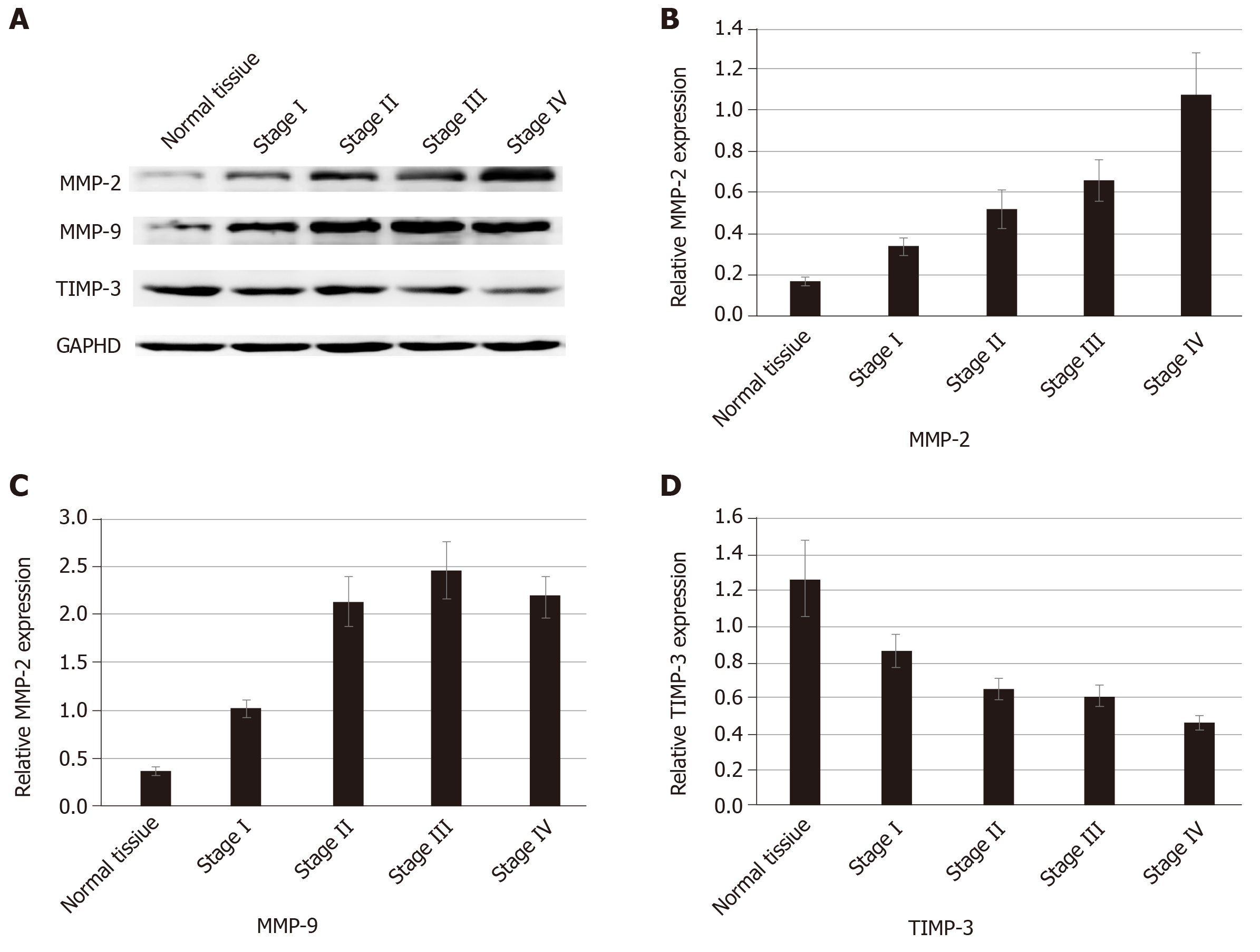
Figure 3 Expression of MMP-2, MMP-9, and TIMP-3 in normal tissue and colorectal cancer.
A: Western blot showing up-regulated expression of MMP-2 and MMP-9 and down-regulated expression of TIMP-3 in colorectal tissues; B: The expression of MMP-2 increased with increased tumor stage; C: The expression of MMP-9 in the colorectal cancer tissues was higher than that in the normal tissue and it was highest in the stage III colorectal cancer; D: The expression of TIMP-3 decreased with increased tumor stage.
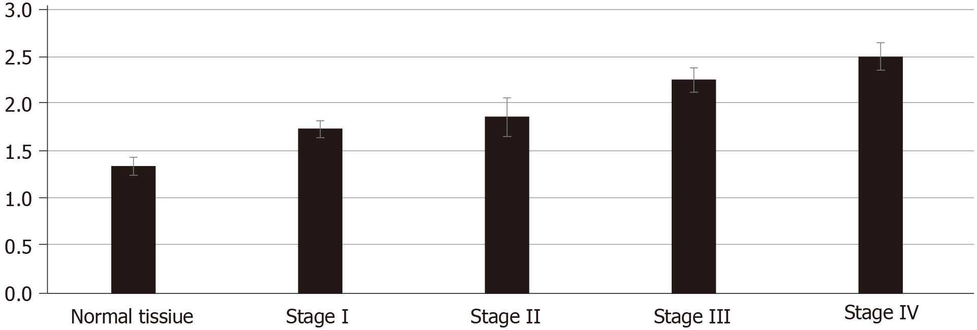
Figure 4 Cancer cells co-cultured with colorectal cancer extracellular matrix and normal tissue extracellular matrix in vitro.
Compared to the optical density (OD) value of the normal tissue extracellular matrix (ECM), the OD value of colorectal cancer (CRC) ECM was higher. When comparing every two OD values of the CRC ECM, the difference between stage I ECM and stage II ECM was not statistically significant (P = 0.138).
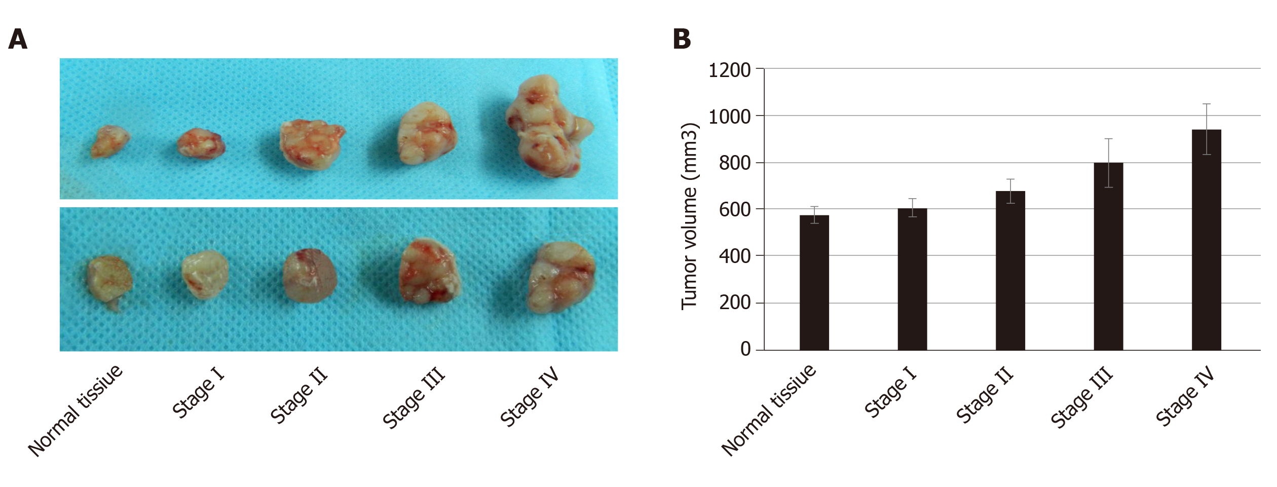
Figure 5 Cancer cells co-cultured with colorectal cancer extracellular matrix and normal tissue extracellular matrix in vivo.
A: The volume of tumor in colorectal cancer extracellular matrix (ECM) was bigger than that in the normal tissue ECM; B: When comparing every two tumor volumes, the differences between stage I ECM and normal tissue ECM and between stage I ECM and stage II ECM were not statistically significant (P = 0.526 and 0.152, respectively).
- Citation: Li ZL, Wang ZJ, Wei GH, Yang Y, Wang XW. Changes in extracellular matrix in different stages of colorectal cancer and their effects on proliferation of cancer cells. World J Gastrointest Oncol 2020; 12(3): 267-275
- URL: https://www.wjgnet.com/1948-5204/full/v12/i3/267.htm
- DOI: https://dx.doi.org/10.4251/wjgo.v12.i3.267


