Copyright
©The Author(s) 2020.
World J Gastrointest Oncol. Jan 15, 2020; 12(1): 77-82
Published online Jan 15, 2020. doi: 10.4251/wjgo.v12.i1.77
Published online Jan 15, 2020. doi: 10.4251/wjgo.v12.i1.77
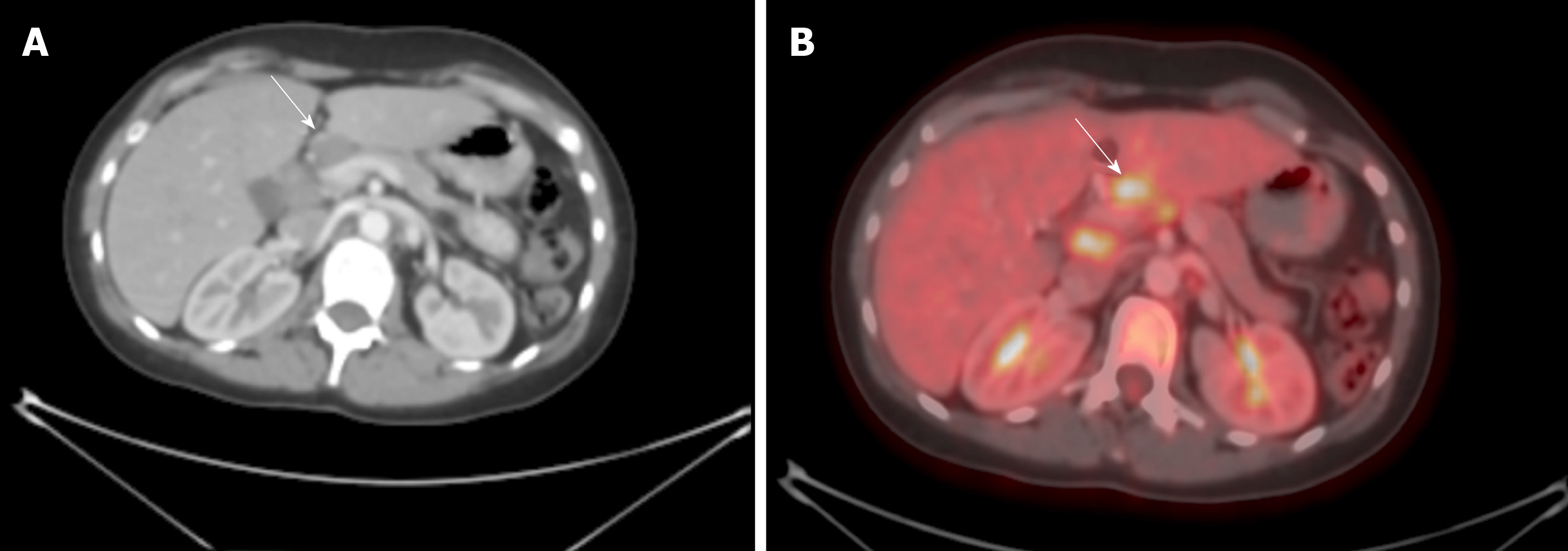
Figure 1 Abnormal FDG uptake on positron emission tomography.
A: Computed tomography scan of the abdomen showing enlarged hepatic LN; B: Positron emission tomography showing abnormal FDG uptake in the same LN. FDG: Fluorodeoxyglucose; LN: Lymph node.
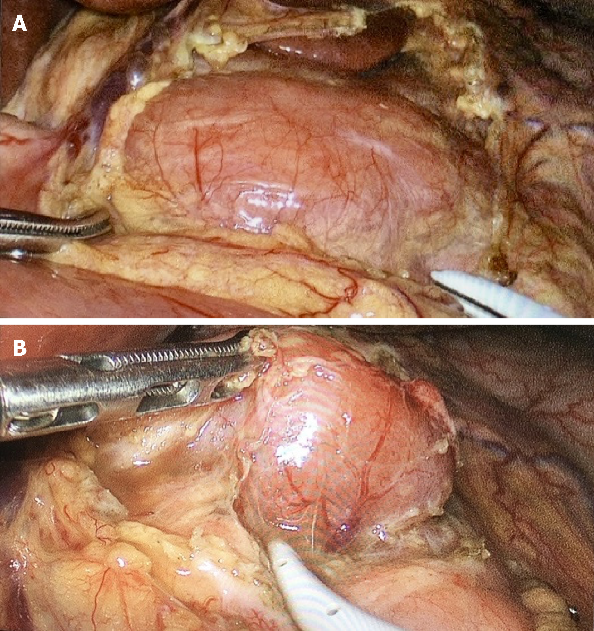
Figure 2 The left lobe of the liver is retracted upwards towards the diaphragm and the lesser omentum with its pars flaccida is exposed.
A: Exposure of the hepatic node excising the lesser omentum; B: Dissection of the hepatic node.
- Citation: Ben-Ishay O. Laparoscopic dissection of the hepatic node: The trans lesser omentum approach. World J Gastrointest Oncol 2020; 12(1): 77-82
- URL: https://www.wjgnet.com/1948-5204/full/v12/i1/77.htm
- DOI: https://dx.doi.org/10.4251/wjgo.v12.i1.77


