Copyright
©The Author(s) 2019.
World J Gastrointest Oncol. Sep 15, 2019; 11(9): 665-678
Published online Sep 15, 2019. doi: 10.4251/wjgo.v11.i9.665
Published online Sep 15, 2019. doi: 10.4251/wjgo.v11.i9.665
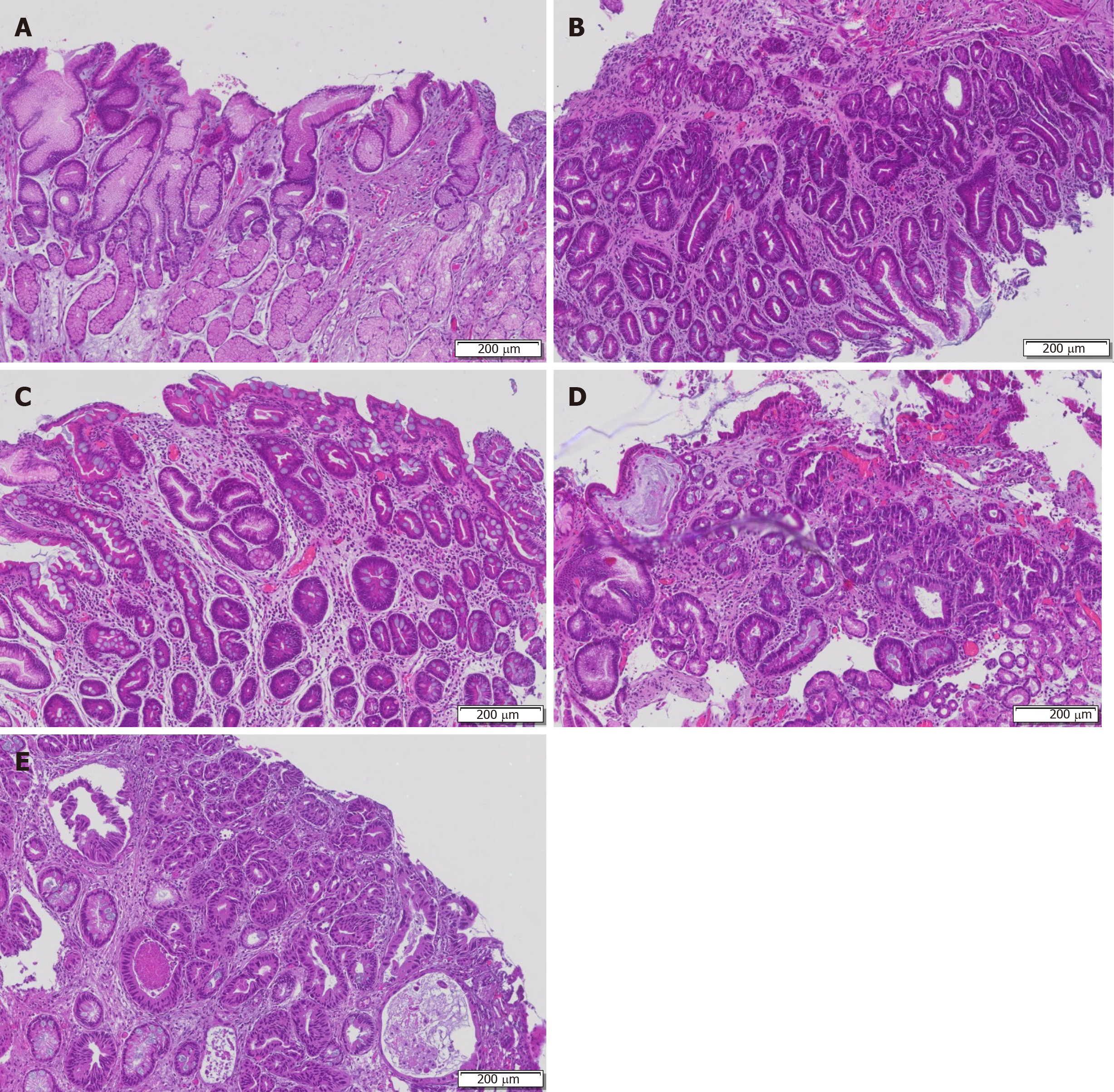
Figure 1 Complete or incomplete subtypes.
A: Chronic gastritis with mucosal atrophy and lymphocytic infiltrate (asterix); B: Incomplete intestinal metaplasia resembling the colonic-type epithelium with irregular mucin droplets (arrowheads) and absence of a brush border; C: Complete intestinal metaplasia resembling the small intestinal epithelium with goblet cells alternating with eosinophilic enterocytes, brush border and Paneth cells; D: Low-grade dysplasia characterized by crowded glands with columnar cells and preserved polarity and pseudostratified nuclei; E: High-grade dysplasia with cuboidal cells, mitotic activity, prominent nucleoi, and high nuclear-cytoplasmia ratio.
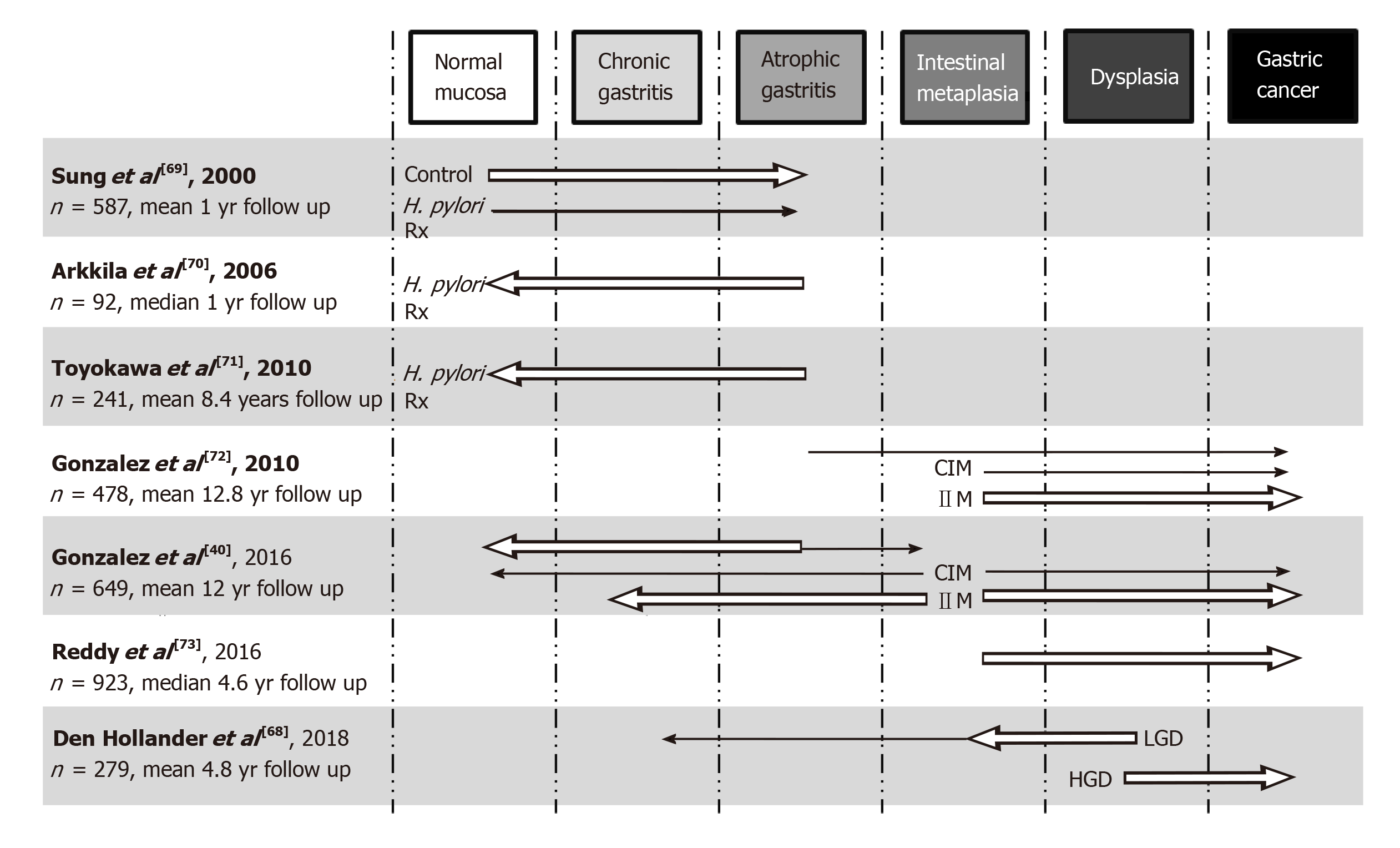
Figure 2 Graphical representation of selected large studies investigating progression/regression of premalignant gastric lesions across the stages of the Correa cascade.
Arrows represent the direction of effect findings, with the size of the arrow the strength of effect (not to scale between cohorts and only major findings of trials represented). H. pylori Rx: Helicobacter pylori antibiotic therapy; CIM: Complete IM; IIM: Incomplete IM; LGD: Low-grade dysplasia; HGD: High-grade dysplasia; IM: Intestinal metaplasia.
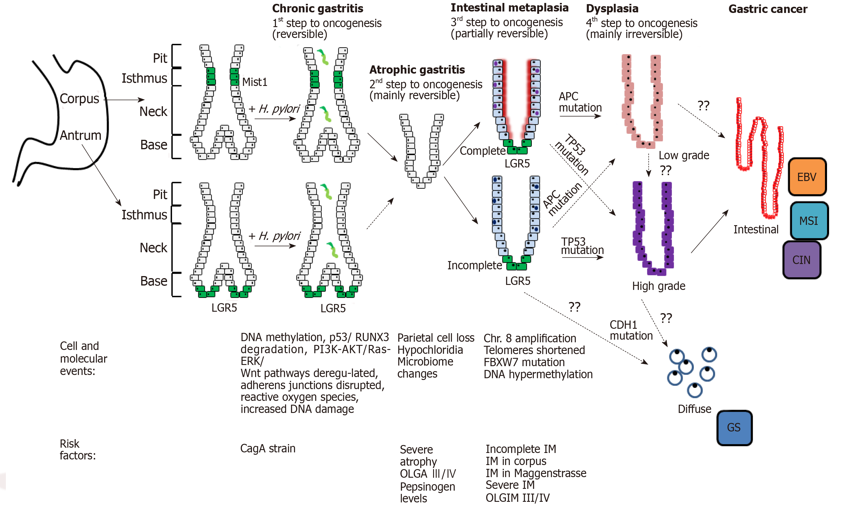
Figure 3 Summary of the cellular and molecular events associated with progression to cancer.
H. pylori: Helicobacter pylori; EBV: Epstein Barr virus positive; MSI: Microsatellite unstable; CagA: Cytotoxin-associated gene A protein; IM: Intestinal metaplasia; GS: Genomically stable.
- Citation: Koulis A, Buckle A, Boussioutas A. Premalignant lesions and gastric cancer: Current understanding. World J Gastrointest Oncol 2019; 11(9): 665-678
- URL: https://www.wjgnet.com/1948-5204/full/v11/i9/665.htm
- DOI: https://dx.doi.org/10.4251/wjgo.v11.i9.665


