Copyright
©The Author(s) 2019.
World J Gastrointest Oncol. May 15, 2019; 11(5): 436-448
Published online May 15, 2019. doi: 10.4251/wjgo.v11.i5.436
Published online May 15, 2019. doi: 10.4251/wjgo.v11.i5.436
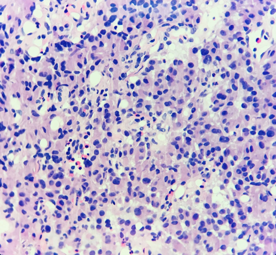
Figure 1 Hematoxylin-eosin staining results of hepatic neuroendocrine neoplasm.
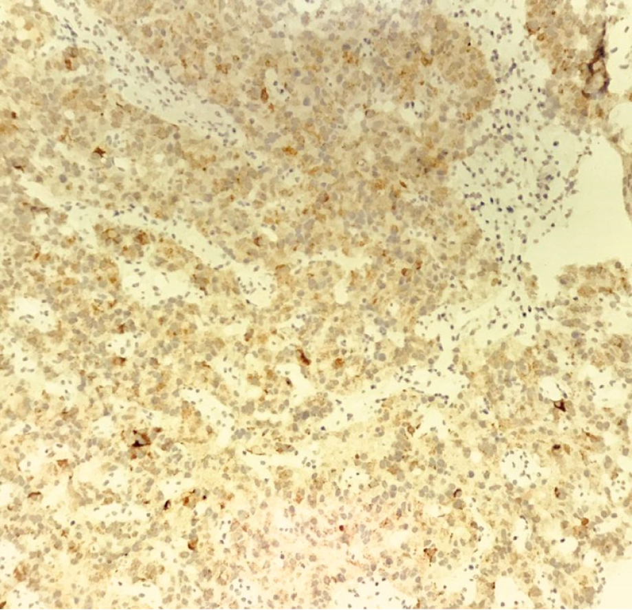
Figure 2 ChrA positive expression in hepatic neuroendocrine neoplasm.
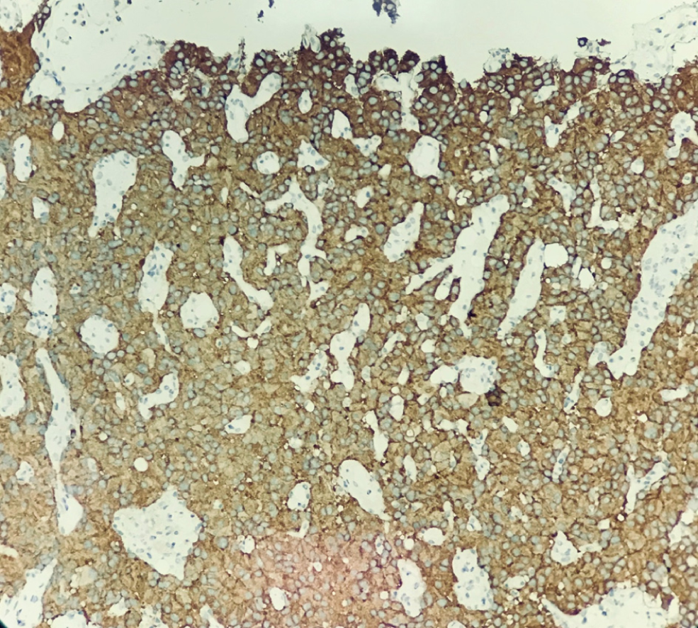
Figure 3 Syno positive expression in hepatic neuroendocrine neoplasm.
- Citation: Kang XN, Zhang XY, Bai J, Wang ZY, Yin WJ, Li L. Analysis of B-ultrasound and contrast-enhanced ultrasound characteristics of different hepatic neuroendocrine neoplasm. World J Gastrointest Oncol 2019; 11(5): 436-448
- URL: https://www.wjgnet.com/1948-5204/full/v11/i5/436.htm
- DOI: https://dx.doi.org/10.4251/wjgo.v11.i5.436


