Copyright
©The Author(s) 2019.
World J Gastrointest Oncol. Feb 15, 2019; 11(2): 172-180
Published online Feb 15, 2019. doi: 10.4251/wjgo.v11.i2.172
Published online Feb 15, 2019. doi: 10.4251/wjgo.v11.i2.172
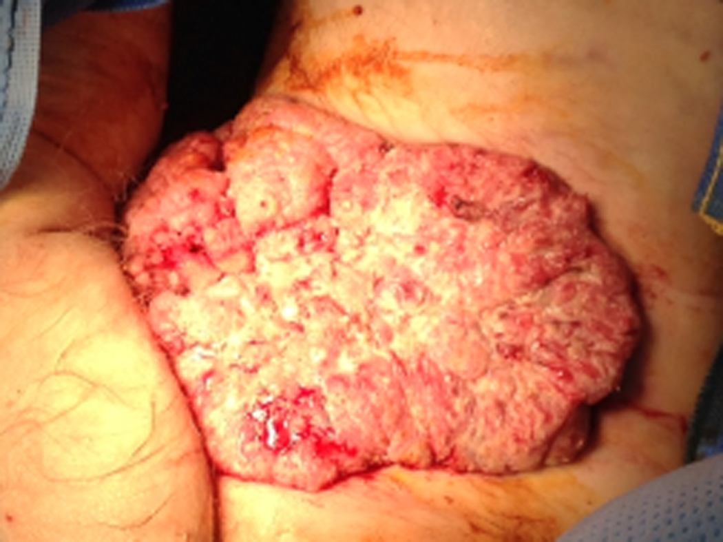
Figure 1 Large anal condyloma arising from anus with the tumor extending into the left scrotal skin, left thigh crease and to the lower left inguinal area (case 1).
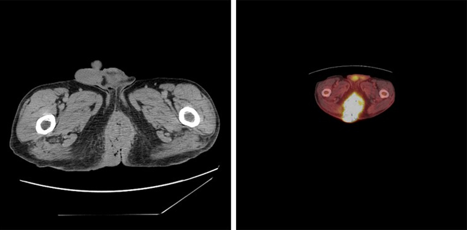
Figure 2 Positron emission tomography computed tomography scan of pelvis showing large perianal, hypermetabolic mass involving the entire left thigh crease, scrotum and the inguinal area (case 1).
Figure 3 Positron emission tomography computed tomography scans of pelvis showing large anal hypermetabolic mass (case 2).
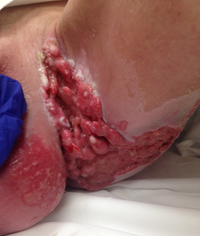
Figure 4 Tumor response 3 mo after the Chemo radiation (Nigro protocol) in case 1.
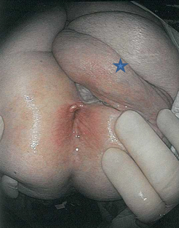
Figure 5 Complete resolution of anal cancer and condyloma, three years follow up.
The blue star denotes the remaining scrotum and VRAM pedicle flap.
- Citation: Shenoy S, Nittala M, Assaf Y. Anal carcinoma in giant anal condyloma, multidisciplinary approach necessary for optimal outcome: Two case reports and review of literature. World J Gastrointest Oncol 2019; 11(2): 172-180
- URL: https://www.wjgnet.com/1948-5204/full/v11/i2/172.htm
- DOI: https://dx.doi.org/10.4251/wjgo.v11.i2.172


