Copyright
©The Author(s) 2018.
World J Gastrointest Oncol. Jul 15, 2018; 10(7): 194-201
Published online Jul 15, 2018. doi: 10.4251/wjgo.v10.i7.194
Published online Jul 15, 2018. doi: 10.4251/wjgo.v10.i7.194
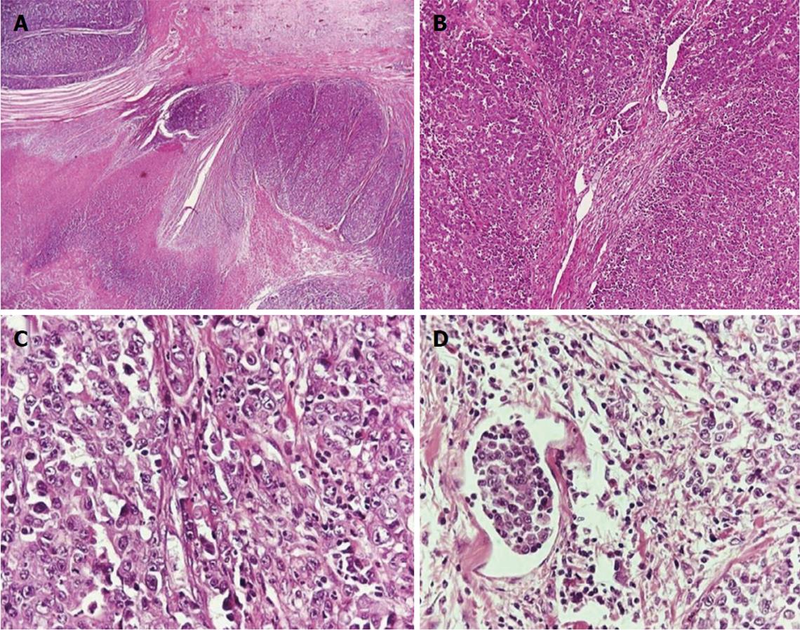
Figure 1 Adenocarcinoma of the small intestine.
A: Low-power view shows the nodular formations of the carcinoma and foci of necrosis (H and E, × 25); B: High-power view shows the diffuse growth pattern and the presence of few tubular structures (H and E, × 100); C: High-power view shows the neoplastic cells with the hyperchromatic, irregular nuclei with prominent nucleoli; D: The presence of lymphatic tumor emboli (H and E, × 400).
Figure 2 Surgical specimen after partial gastrectomy of the gastric pouch with resection of the Braun anastomosis.
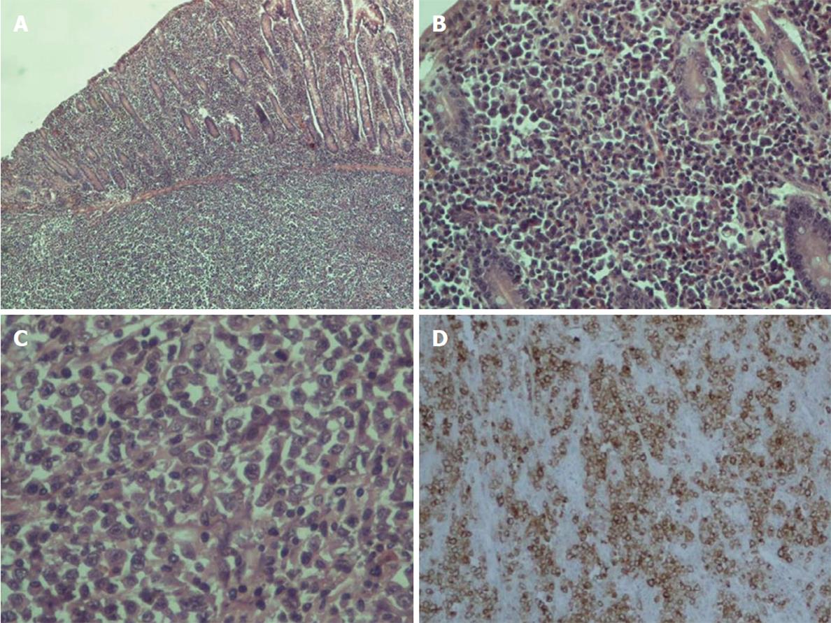
Figure 3 Anaplastic large cell lymphoma of the small bowel.
A: H and E stain × 40; B: H and E stain × 200; C: H and E stain × 400; D: Ki-1 antigen (CD30) staining (+).
- Citation: Kotidis E, Ioannidis O, Pramateftakis MG, Christou K, Kanellos I, Tsalis K. Atypical anastomotic malignancies of small bowel after subtotal gastrectomy with Billorth II gastroenterostomy for peptic ulcer: Report of three cases and review of the literature. World J Gastrointest Oncol 2018; 10(7): 194-201
- URL: https://www.wjgnet.com/1948-5204/full/v10/i7/194.htm
- DOI: https://dx.doi.org/10.4251/wjgo.v10.i7.194


