Copyright
©The Author(s) 2018.
World J Gastrointest Oncol. Jun 15, 2018; 10(6): 137-144
Published online Jun 15, 2018. doi: 10.4251/wjgo.v10.i6.137
Published online Jun 15, 2018. doi: 10.4251/wjgo.v10.i6.137
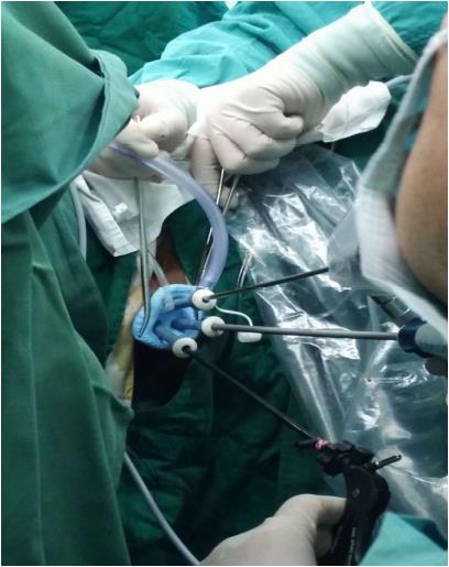
Figure 1 Settings for trans-anal minimally invasive surgery.
The SILS-Port® was inserted through the anus. Assisting trocars and routine laparoscopic instruments were placed. A high-definition 30º 5 mm or 10 mm laparoscopic camera lens was chosen. SILS: Single incision laparoscopic surgery.
Figure 2 Procedures of trans-anal minimally invasive surgery.
A: Intra-operative view of TAMIS showed resection margin was marked by electrocautery; B: An endoscopic grasper and electrocautery were used to facilitate a fullthickness excision; C: The defect of rectal wall was closed using STRATAFIXTM; D: The surgical specimen was pinned on plastic board with indicative orientation. TAMIS: Trans-anal minimally invasive surgery.
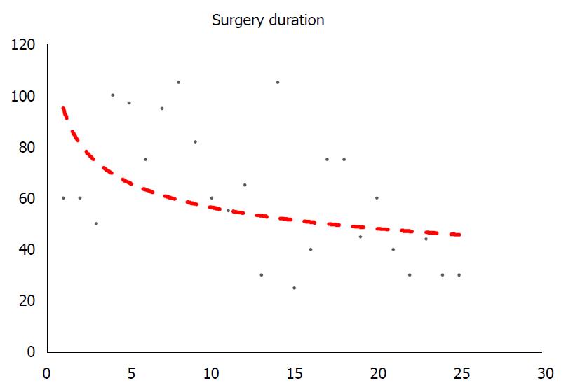
Figure 3 Correlation between cases and duration of trans-anal minimally invasive surgery surgeries.
The X-axis represented individual case consequently, while the Y-axis was the duration of each surgery (min), indicating the learning curve of this technique.
- Citation: Chen N, Peng YF, Yao YF, Gu J. Trans-anal minimally invasive surgery for rectal neoplasia: Experience from single tertiary institution in China. World J Gastrointest Oncol 2018; 10(6): 137-144
- URL: https://www.wjgnet.com/1948-5204/full/v10/i6/137.htm
- DOI: https://dx.doi.org/10.4251/wjgo.v10.i6.137


