Copyright
©The Author(s) 2018.
World J Gastrointest Oncol. Dec 15, 2018; 10(12): 522-527
Published online Dec 15, 2018. doi: 10.4251/wjgo.v10.i12.522
Published online Dec 15, 2018. doi: 10.4251/wjgo.v10.i12.522
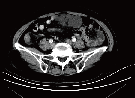
Figure 1 Abdominal computed tomography scan reveals lumpy low-density shadows around the upper middle intestine, 86 mm × 42 mm in size, and no enhancement was observed (Case 4).
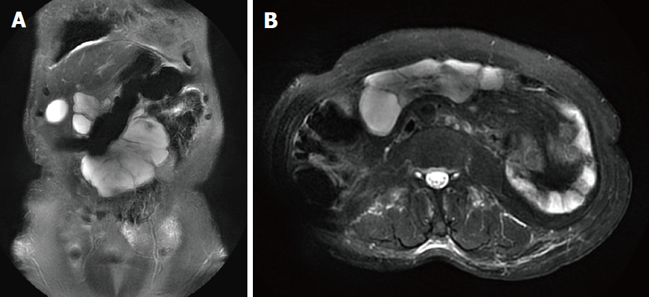
Figure 2 Abdominal magnetic resonance imaging shows cystic long T1 and long T2 signal masses in the anterior wall of the middle abdomen, 110 mm × 40 mm in size.
No abnormal enhancement was observed (Case 4). A: Coronal position; B: Axial position.
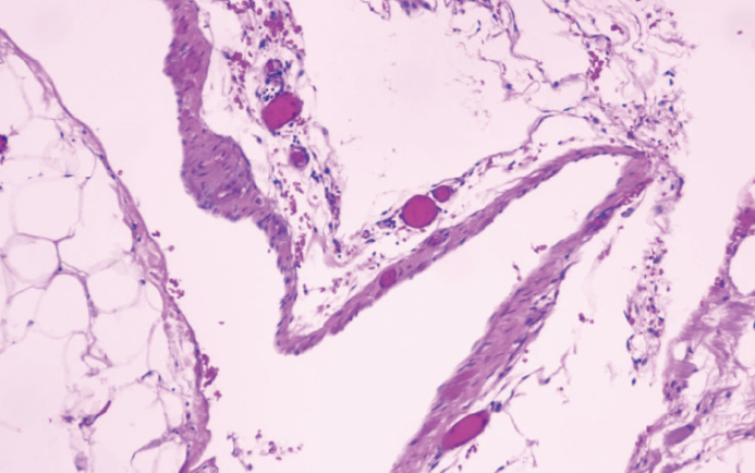
Figure 3 Histopathology shows that the cyst wall was composed of fibrous adipose tissue and a few lymphatic endothelial proliferations.
Case 4, hematoxylin and eosin, 100 ×.
- Citation: Chen J, Du L, Wang DR. Experience in the diagnosis and treatment of mesenteric lymphangioma in adults: A case report and review of literature. World J Gastrointest Oncol 2018; 10(12): 522-527
- URL: https://www.wjgnet.com/1948-5204/full/v10/i12/522.htm
- DOI: https://dx.doi.org/10.4251/wjgo.v10.i12.522


