Copyright
©2009 Baishideng.
World J Gastrointest Oncol. Oct 15, 2009; 1(1): 93-96
Published online Oct 15, 2009. doi: 10.4251/wjgo.v1.i1.93
Published online Oct 15, 2009. doi: 10.4251/wjgo.v1.i1.93
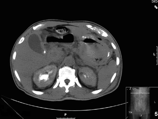
Figure 1 CAT scan of the abdomen revealed thickening and calcification of the gastric wall with associated pneumatosis, as well as a 1 cm pedunculated mass in the proximal duodenum.
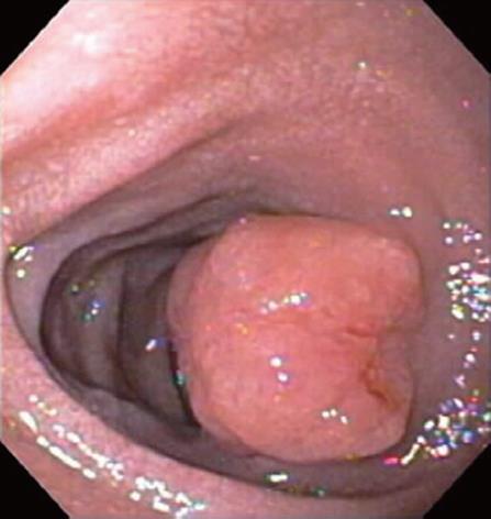
Figure 2 On esophagogastroduodenoscopy, a non bleeding 3 cm polyp was noted in the post-bulbar area of the duodenum.
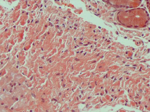
Figure 3 Under light microscopy, gastric biopsy appears salmon-colored when stained with Congo Red.
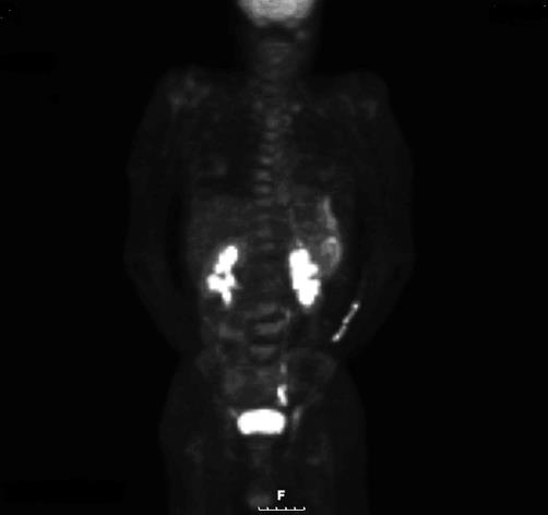
Figure 4 PET scan revealed diffuse gastric mucosa uptake compatible with gastric malignancy but no hypermetabolic neoplastic foci elsewhere.
- Citation: Koczka CP, Goodman AJ. Gastric amyloidoma in patient after remission of Non-Hodgkin’s Lymphoma. World J Gastrointest Oncol 2009; 1(1): 93-96
- URL: https://www.wjgnet.com/1948-5204/full/v1/i1/93.htm
- DOI: https://dx.doi.org/10.4251/wjgo.v1.i1.93


