Copyright
©2009 Baishideng.
World J Gastrointest Oncol. Oct 15, 2009; 1(1): 55-61
Published online Oct 15, 2009. doi: 10.4251/wjgo.v1.i1.55
Published online Oct 15, 2009. doi: 10.4251/wjgo.v1.i1.55
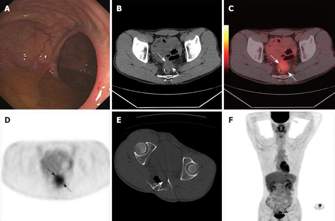
Figure 1 Perirectal recurrence.
Endoscopic examination, transverse images and PET/CT images obtained 25 mo after surgery in a 39-year-old man. A: Colonoscopy at the level of tumor resection showed no evidence of recurrence; B: CT demonstrated perirectal soft tissue that might represent a local recurrent tumor or postoperative scar tissue (arrows); C, D: PET/CT revealed a perirectal lesion with high FDG uptake (arrows); E: Local recurrence was confirmed by PET/CT guided tissue core biopsy (arrow); F: Whole body PET/CT confirmed local recurrent tumor (arrow) and no distant metastasis.
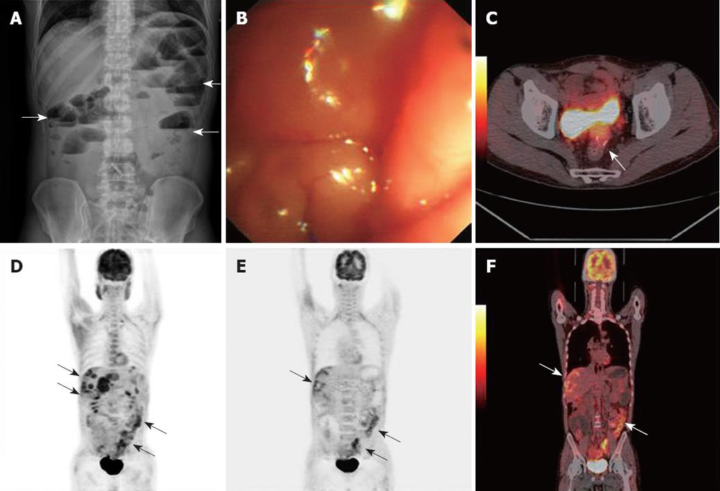
Figure 2 Extrapelvic metastases and perirectal recurrence.
Plain abdominal radiograph, ES and PET/CT images obtained 30 mo after rectal cancer resection in a 41-year-old man. A, B: Plain abdominal radiograph and colonoscopy revealed the obstruction at the anastomotic site (arrows); C-F: PET/CT showed perirectal recurrence, peritoneal carcinomatosis and liver metastases (arrows).
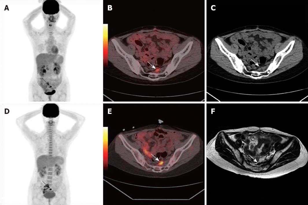
Figure 3 False-positive results.
PET/CT obtained in a 34-year-old woman 16 mo after rectal cancer resection. A-C: 18F-FDG uptake in the left side of the pelvis was misinterpreted as tumor recurrence (arrows); D, E: 18F-FDG uptake in the left side of the pelvis was unchanged after 6 mo PET/CT scan (arrows); F: MRI showed no changes of lesion 11 mo follow-up after first PET/CT examination (arrow). The final diagnosis of scar tissue was established by clinical and imaging follow-up.
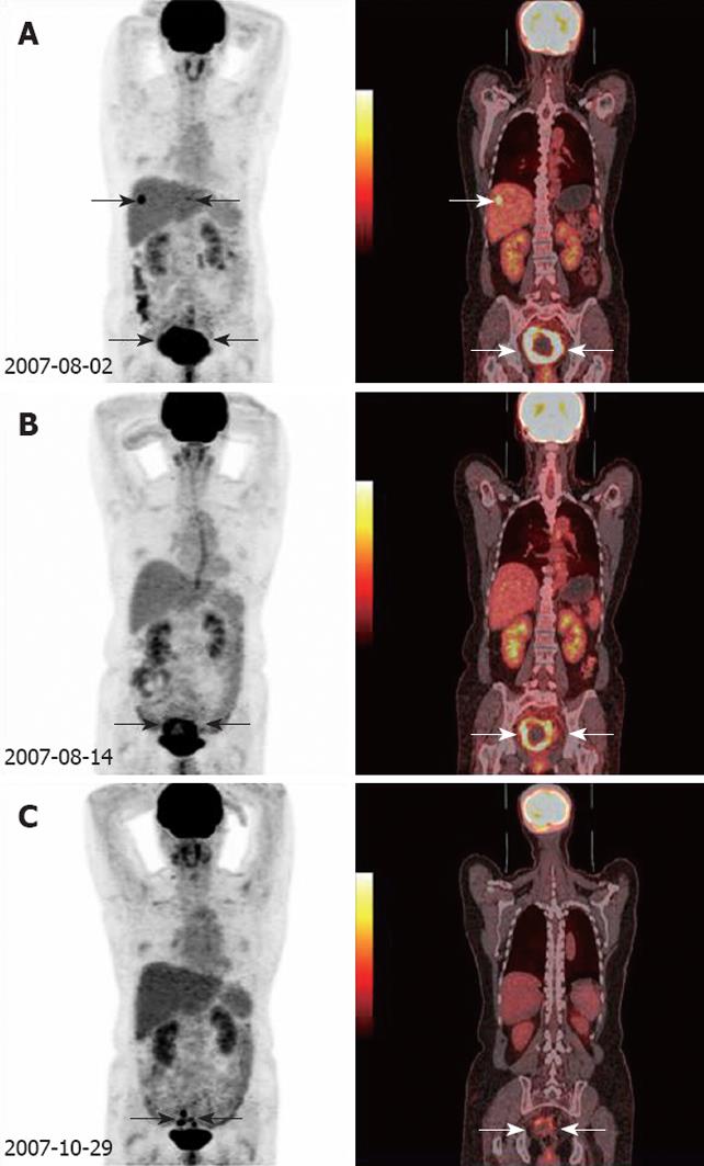
Figure 4 Monitoring responses to pelvic recurrence and liver metastases.
Three PET/CT scans were obtained in a 55-year-old woman five months after rectal cancer resection. A: PET/CT demonstrated a locoregional recurrence and liver metastasis foci (arrows) before salvage treatment; B: PET/CT showed obvious decreasing of intense activity after two cycles of chemotherapy (arrows); C: After six cycles of chemotherapy, PETCT imaging follow-up revealed good control of pelvic recurrence and liver metastases (arrows).
- Citation: Sun L, Guan YS, Pan WM, Luo ZM, Wei JH, Zhao L, Wu H. Clinical value of 18F-FDG PET/CT in assessing suspicious relapse after rectal cancer resection. World J Gastrointest Oncol 2009; 1(1): 55-61
- URL: https://www.wjgnet.com/1948-5204/full/v1/i1/55.htm
- DOI: https://dx.doi.org/10.4251/wjgo.v1.i1.55


