Published online Sep 16, 2013. doi: 10.4253/wjge.v5.i9.465
Revised: June 19, 2013
Accepted: July 30, 2013
Published online: September 16, 2013
Processing time: 124 Days and 17.4 Hours
In capsule endoscopy (CE), there is research to develop hardware that enables ‘‘real’’ three-dimensional (3-D) video. However, it should not be forgotten that ‘‘true’’ 3-D requires dual video images. Inclusion of two cameras within the shell of a capsule endoscope though might be unwieldy at present. Therefore, in an attempt to approximate a 3-D reconstruction of the digestive tract surface, a software that recovers information-using gradual variation of shading-from monocular two-dimensional CE images has been proposed. Light reflections on the surface of the digestive tract are still a significant problem. Therefore, a phantom model and simulator has been constructed in an attempt to check the validity of a highlight suppression algorithm. Our results confirm that 3-D representation software performs better with simultaneous application of a highlight reduction algorithm. Furthermore, 3-D representation follows a good approximation of the real distance to the lumen surface.
Core tip: In an attempt to approximate a three-dimensional (3-D) reconstruction of the digestive tract surface, a software that recovers information-using gradual variation of shading - from monocular two-dimensional capsule endoscopy images has been proposed. Light reflections on the surface of the digestive tract are still a significant problem. Therefore, a phantom model and simulator has been constructed in an attempt to check the validity of a highlight suppression algorithm. Our results confirm that 3-D representation software performs better with simultaneous application of a highlight reduction algorithm. Furthermore, 3-D representation follows a good approximation of the real distance to the lumen surface.
- Citation: Koulaouzidis A, Karargyris A. Use of enhancement algorithm to suppress reflections in 3-D reconstructed capsule endoscopy images. World J Gastrointest Endosc 2013; 5(9): 465-467
- URL: https://www.wjgnet.com/1948-5190/full/v5/i9/465.htm
- DOI: https://dx.doi.org/10.4253/wjge.v5.i9.465
In capsule endoscopy (CE), there is research to develop hardware that enables ‘‘real’’ three-dimensional (3-D) video by using an infrared projector and a CMOS camera[1,2]. However, it should not be forgotten that ‘‘true’’ 3-D requires dual video-images; furthermore, the inclusion of two cameras within the shell of a capsule endoscope might be unwieldy at present[3]. Therefore, major drawbacks at present are size, power consumption and packaging issues[4]. In an attempt to approximate a 3-D reconstruction of the digestive tract surface, Koulaouzidis et al[4] and Karargyris et al[5] proposed the use of a software [Shape-from-Shading (SfS)] that utilizes monocular CE frames. Essentially, SfS algorithms recover information -using gradual variation of shading[6]- on the shape of objects given a single two-dimensional (2-D) image. 3-D representation may be helpful in conjunction with other image enhancement tools e.g., virtual chromoendoscopy (FICE)[7] and/or color (blue) mode analysis of CE videos[8].
However, light reflections on the surface of the digestive tract are still a significant problem, not only for 3-D representation but also for traditional 2-D CE. When light falls on to a surface, some of the beams are reflected back straightaway -specular reflection- while the rest of the beams penetrate it before reflected (diffuse reflection). As most digestive tract structures/surfaces are di-electric and homogeneous, they display both types of reflections[4]. To reduce reflections, a highlight suppression algorithm[9] has been applied onto CE images.
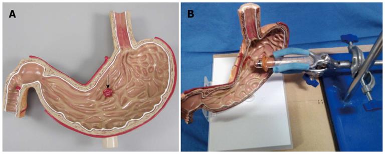
To test this algorithm, a phantom task simulator was created. A Stomach Ulcer Anatomical Model (manufacturer: Anatomical Chart Company G200) was used; the stomach model has an red-colored base ulcer (1/2”diameter and 3/16”depth; Figure 1A); the latter was thereafter colored buttercup yellow using quick-drying spray paint (Tor Coatings®Ltd., United Kingdom) and white (using flat white spray from Plasti-Kote®Ltd.). A PillCam®SB2 (Given®Imaging Ltd., Yoqneam, Israel) was mounted on a plastic tube and held (with the use of regular lab stand) at 0, 5, 10, 15 and 20 mm from the ulcer base (usual working distance of the CE in vivo, Figure 1B). The images were uploaded to a workstation and they were categorized based on distance and ulcer base color (red, yellow and white). We aimed to check whether the ulcer models appear closer or further based on their 3-D representation.
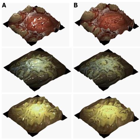
Tsai’s SfS[9,10] algorithm was applied on each image in order to reconstruct its 3-D representation with (Figure 2A) or without (Figure 2B) software highlight suppression[9]. Tsai’s SfS algorithm cannot measure the real distance of the camera to the model’s surface but it gives the relative distance (z) to the black frame background. For each image, we selected the region of interest (ROI) of the ulcer model on the 3-D representation and we calculated the average depth (z) for each ROI.
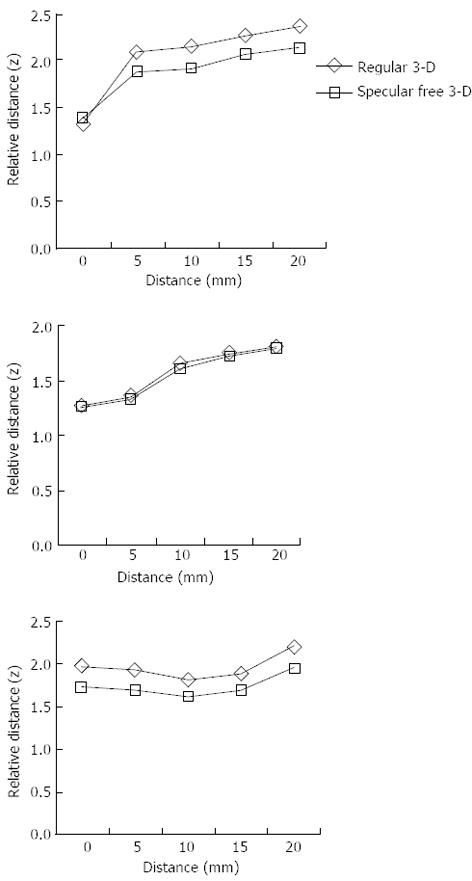
The results (charts, Figure 3) confirm that the distance of the camera from the model surface increases so does the relative distance (z) on the 3-D representation. This effect is more evident for the white and yellow ulcer models. However, relative distance does not follow a similar trend for the red-based ulcer model. This is likely due to the saturation of the red color creating variations to the shading: red color appears darker or lighter. Finally, from the charts we conclude that the highlight suppression algorithm improved the quality of the images.
In conclusion, 3-D representation software seems to perform better with simultaneous application of a highlight reduction algorithm. Furthermore, 3-D representation follows a good approximation of the real distance to the lumen surface.
P- Reviewers Calabrese C, Riccioni ME S- Editor Zhai HH L- Editor A E- Editor Wang CH
| 1. | Available from: http: //www.visionsense.com/Stereo_Vision.html. |
| 2. | Kolar A, Romain O, Ayoub J, Viateur S, Granado B. Prototype of video endoscopic capsule with 3-d imaging capabilities. IEEE Trans Biomed Circuits Syst. 2010;4:239-249. [RCA] [PubMed] [DOI] [Full Text] [Cited by in Crossref: 21] [Cited by in RCA: 14] [Article Influence: 0.9] [Reference Citation Analysis (0)] |
| 3. | Fisher LR, Hasler WL. New vision in video capsule endoscopy: current status and future directions. Nat Rev Gastroenterol Hepatol. 2012;9:392-405. [RCA] [PubMed] [DOI] [Full Text] [Cited by in Crossref: 50] [Cited by in RCA: 56] [Article Influence: 4.3] [Reference Citation Analysis (0)] |
| 4. | Koulaouzidis A, Karargyris A. Three-dimensional image reconstruction in capsule endoscopy. World J Gastroenterol. 2012;18:4086-4090. [RCA] [PubMed] [DOI] [Full Text] [Full Text (PDF)] [Cited by in CrossRef: 15] [Cited by in RCA: 16] [Article Influence: 1.2] [Reference Citation Analysis (0)] |
| 5. | Karargyris A, Bourbakis N. Three-dimensional reconstruction of the digestive wall in capsule endoscopy videos using elastic video interpolation. IEEE Trans Med Imaging. 2011;30:957-971. [RCA] [PubMed] [DOI] [Full Text] [Cited by in Crossref: 46] [Cited by in RCA: 26] [Article Influence: 1.9] [Reference Citation Analysis (0)] |
| 6. | Zhang R, Tsai PS, Cryer JE, Shah M. Shape from Shading: A Survey. IEEE Trans Pattern Anal Mach Intell. 1999;21:690-706. [DOI] [Full Text] |
| 7. | Krystallis C, Koulaouzidis A, Douglas S, Plevris JN. Chromoendoscopy in small bowel capsule endoscopy: Blue mode or Fuji Intelligent Colour Enhancement? Dig Liver Dis. 2011;43:953-957. [RCA] [PubMed] [DOI] [Full Text] [Cited by in Crossref: 24] [Cited by in RCA: 33] [Article Influence: 2.4] [Reference Citation Analysis (0)] |
| 8. | Koulaouzidis A, Douglas S, Plevris JN. Blue mode does not offer any benefit over white light when calculating Lewis score in small-bowel capsule endoscopy. World J Gastrointest Endosc. 2012;4:33-37. [RCA] [PubMed] [DOI] [Full Text] [Full Text (PDF)] [Cited by in CrossRef: 18] [Cited by in RCA: 24] [Article Influence: 1.8] [Reference Citation Analysis (0)] |
| 9. | Tan RT, Ikeuchi K. Separating reflection components of textured surfaces using a single image. IEEE Trans Pattern Anal Mach Intell. 2005;27:178-193. [PubMed] [DOI] [Full Text] |
| 10. | Koulaouzidis A, Karargyris A, Rondonotti E, Noble CL, Douglas S, Alexandridis E, Zahid AM, Bathgate AJ, Trimble KC, Plevris JN. Three-dimensional representation software as image enhancement tool in small-bowel capsule endoscopy: A feasibility study. Dig Liver Dis. 2013;Epub ahead of print. [RCA] [PubMed] [DOI] [Full Text] [Cited by in Crossref: 16] [Cited by in RCA: 23] [Article Influence: 1.9] [Reference Citation Analysis (0)] |


