Published online Apr 16, 2013. doi: 10.4253/wjge.v5.i4.189
Revised: November 5, 2012
Accepted: January 5, 2013
Published online: April 16, 2013
Processing time: 197 Days and 5.9 Hours
Ascaris lumbricoides (A. lumbricoides) is the most common intestinal roundworm parasite, infecting approximately one quarter of the world’s population. Infection can lead to various complications because it can spread along the gastrointestinal tract. Although A. lumbricoides infection is a serious healthcare issue in developing countries, it now also has a worldwide distribution as a result of increased immigration and travel. Intestinal obstruction is the most common complication of A. lumbricoides infection, potentially leading to even more serious consequences such as small bowel perforation and peritonitis. Diagnosis is based primarily on stool samples and the patient’s history. Early diagnosis, aided in part by knowledge of the local prevalence, can result in early treatment, thereby preventing surgical complications associated with intestinal obstruction. Further, delay in diagnosis may have fatal consequences. Capsule endoscopy can serve as a crucial, non-invasive diagnostic tool for A. lumbricoides infection, especially when other diagnostic methods have failed to detect the parasite. We report a case of A. lumbricoides infection that resulted in intestinal obstruction at the level of the ileum. Both stool sample examination and open surgery failed to indicate the presence of A. lumbricoides, and the cause of the obstruction was only revealed by capsule endoscopy. The patient was treated with anthelmintics.
-
Citation: Yamashita ET, Takahashi W, Kuwashima DY, Langoni TR, Costa-Genzini A. Diagnosis of
Ascaris lumbricoides infection using capsule endoscopy. World J Gastrointest Endosc 2013; 5(4): 189-190 - URL: https://www.wjgnet.com/1948-5190/full/v5/i4/189.htm
- DOI: https://dx.doi.org/10.4253/wjge.v5.i4.189
Ascaris lumbricoides (A. lumbricoides) has a worldwide distribution, but occurs most frequently in underdeveloped regions where sanitation is poor[1,2]. In most cases the infection remains asymptomatic until the number of worms in the intestines increases considerably. It can cause serious complications, the most common of which is intestinal obstruction, although pancreatitis, cholangitis, bleeding, and obstructive jaundice can also occur[3,4]. The diagnosis of A. lumbricoides infection is based mainly on patient history and stool samples, but complementary exams such as abdominal radiography and computed tomography can also aid in the diagnosis[5]. We report a case of A. lumbricoides infection that resulted in intestinal obstruction. Although the obstruction was apparent during open surgery and imaging, neither they, nor the stool samples analysis revealed the presence of A. lumbricoides. The presence of this parasite was however determined by video capsule endoscopy.
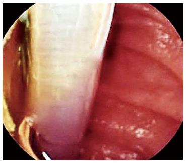
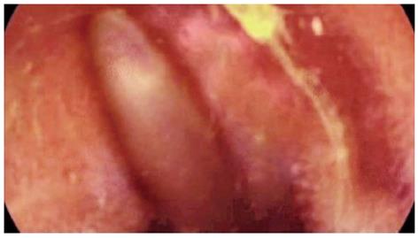
A 64-year-old Brazilian woman presented with abdominal discomfort and intermittent subocclusive episodes that had developed over the previous few weeks. The discomfort was relieved by evacuation. Physical examination indicated good health, and no abdominal tenderness was noted. The patient had undergone 2 previous exploratory laparoscopy procedures to examine the subocclusion, but the findings were normal. A stool sample was analyzed to detect the possible presence of a parasitic infection, but the findings were negative. However, contrast radiography and computed tomography revealed a partial obstruction with an undetermined tube-like structure at the level of the ileum, suggesting a parasitic infection. Capsule endoscopy (MiroCam capsule; Intromedic, Seoul, South Korea) was performed to determine the cause of the obstruction. A diagnosis of roundworm infection with partial obstruction of the ileum with live A. lumbricoides was confirmed (Figures 1 and 2). The first roundworm was seen 1 h 34 min after capsule ingestion (Figure 1) and the last one was seen 2 h later (Figure 2). Treatment with albendazole and piperazine was initiated, and the patient made a full recovery.
A. lumbricoides is the most common intestinal helminth parasite, infecting approximately one quarter of the world’s population[6]. It has long been endemic in developing countries, but it now has a worldwide distribution due to the increase in immigration and travel[7]. Capsule endoscopy is an important tool for evaluation of small bowel disorders, allowing for non-invasive diagnosis of many diseases. In this case, it was used successfully to reveal the cause of intestinal obstruction as being due to A. lumbricoides infection. This was after stool sample analysis and open surgery, which are currently considered to be the gold standard for A. lumbricoides.
P- Reviewer Tokuhara D S- Editor Song XX L- Editor A E- Editor Zhang DN
| 1. | Cooper PJ, Chico ME, Sandoval C, Espinel I, Guevara A, Kennedy MW, Urban Jr JF, Griffin GE, Nutman TB. Human infection with Ascaris lumbricoides is associated with a polarized cytokine response. J Infect Dis. 2000;182:1207-1213. [RCA] [PubMed] [DOI] [Full Text] [Cited by in Crossref: 184] [Cited by in RCA: 173] [Article Influence: 6.9] [Reference Citation Analysis (0)] |
| 2. | Ziegelbauer K, Speich B, Mäusezahl D, Bos R, Keiser J, Utzinger J. Effect of sanitation on soil-transmitted helminth infection: systematic review and meta-analysis. PLoS Med. 2012;9:e1001162. [RCA] [PubMed] [DOI] [Full Text] [Full Text (PDF)] [Cited by in Crossref: 389] [Cited by in RCA: 348] [Article Influence: 26.8] [Reference Citation Analysis (0)] |
| 3. | Akgun Y. Intestinal obstruction caused by Ascaris lumbricoides. Dis Colon Rectum. 1996;39:1159-1163. [RCA] [PubMed] [DOI] [Full Text] [Cited by in Crossref: 21] [Cited by in RCA: 23] [Article Influence: 0.8] [Reference Citation Analysis (0)] |
| 4. | de Silva NR, Guyatt HL, Bundy DA. Morbidity and mortality due to Ascaris-induced intestinal obstruction. Trans R Soc Trop Med Hyg. 1997;91:31-36. [RCA] [PubMed] [DOI] [Full Text] [Cited by in Crossref: 91] [Cited by in RCA: 64] [Article Influence: 2.3] [Reference Citation Analysis (0)] |
| 5. | Reeder MM. The radiological and ultrasound evaluation of ascariasis of the gastrointestinal, biliary, and respiratory tracts. Semin Roentgenol. 1998;33:57-78. [RCA] [PubMed] [DOI] [Full Text] [Cited by in Crossref: 18] [Cited by in RCA: 18] [Article Influence: 0.7] [Reference Citation Analysis (0)] |
| 6. | Zheng PP, Wang BY, Wang F, Ao R, Wang Y. Esophageal space-occupying lesion caused by Ascaris lumbricoides. World J Gastroenterol. 2012;18:1552-1554. [RCA] [PubMed] [DOI] [Full Text] [Full Text (PDF)] [Cited by in CrossRef: 8] [Cited by in RCA: 12] [Article Influence: 0.9] [Reference Citation Analysis (0)] |
| 7. | Masucci L, Graffeo R, Bani S, Bugli F, Boccia S, Nicolotti N, Fiori B, Fadda G, Spanu T. Intestinal parasites isolated in a large teaching hospital, Italy, 1 May 2006 to 31 December 2008. Euro Surveill. 2011;16:19891. [PubMed] |


