Case Report Open Access
Copyright ©The Author(s) 2024. Published by Baishideng Publishing Group Inc. All rights reserved.
World J Gastrointest Endosc. Jun 16, 2024; 16(6): 361-367
Published online Jun 16, 2024. doi: 10.4253/wjge.v16.i6.361
Pleomorphic leiomyosarcoma of the maxilla with metastasis to the colon: A case report
Asma Alnajjar, Abdulrahman Alfadda, Abdullah Mohammad Alqaraawi, Bader Alajlan, Section of Gastroenterology, Department of Medicine, King Faisal Specialist Hospital & Research Centre, Riyadh 11211, Saudi Arabia
Jean Paul Atallah, Department of Oncology, King Faisal Specialist Hospital & Research Centre, Riyadh 11211, Saudi Arabia
Hussah Fahad AlHussaini, Department of Pathology, King Faisal Specialist Hospital & Research Centre, Riyadh 11211, Saudi Arabia
ORCID number: Abdulrahman Alfadda (0009-0002-2952-6957); Abdullah Mohammad Alqaraawi (0000-0003-3800-4827).
Author contributions: Alqaraawi AM, Alfadda A, and Alajlan B proposed the idea of reporting this case; Alqaraawi AM and Alnajjar A were responsible for performing colonoscopy; Alfadda A assisted in removing the ascending colon polyp; Atallah JP was the patient’s primary oncologist; Atallah JP and Alnajjar A collected the data; Alhussaini HF provided pathology slides with reports of the findings; Alnajjar A was responsible for writing the manuscript; Alfadda A, Alqaraawi AM, and Alajlan B were responsible for revising and editing the manuscript, conceptualization, and supervision; and all authors have reviewed the literature review, and read and approved the final manuscript.
Informed consent statement: Informed written consent was obtained from the patients for the publication of this report and any accompanying images.
Conflict-of-interest statement: The authors declare that they have no conflicts of interest to disclose.
CARE Checklist (2016) statement: The authors have read the CARE Checklist (2016), and the manuscript was prepared and revised according to the CARE Checklist (2016).
Open-Access: This article is an open-access article that was selected by an in-house editor and fully peer-reviewed by external reviewers. It is distributed in accordance with the Creative Commons Attribution NonCommercial (CC BY-NC 4.0) license, which permits others to distribute, remix, adapt, build upon this work non-commercially, and license their derivative works on different terms, provided the original work is properly cited and the use is non-commercial. See: https://creativecommons.org/Licenses/by-nc/4.0/
Corresponding author: Abdulrahman Alfadda, FRCP (C), MBBS, MD, Associate Professor, Doctor, Section of Gastroenterology, Department of Medicine, King Faisal Specialist Hospital & Research Centre, Al Mathar Ash Shamali, Riyadh 11211, Saudi Arabia. abalfadda@gmail.com
Received: March 16, 2024
Revised: April 26, 2024
Accepted: May 17, 2024
Published online: June 16, 2024
Processing time: 89 Days and 14.3 Hours

Abstract
BACKGROUND

Pleomorphic leiomyosarcomas make up around 8.6% of all leiomyosarcomas. They behave aggressively and often have poor prognoses. They can affect the gastrointestinal tract and retroperitoneum. To date, pleomorphic leiomyosarcoma involving the mesocolon have been reported in nine patients.

CASE SUMMARY

The patient was a 44-year-old man with a history of pleomorphic leiomyosarcoma of the left maxilla with metastasis to the lung and liver. His most recent positron emission tomography-computed tomography (PET-CT) scan showed uptake in the ascending and transverse colons. A colonoscopy revealed a 5.0 cm × 3.5 cm × 3.0 cm pedunculated polyp in the ascending colon. The polyp was removed using hot snare polypectomy technique and retrieved with Rothnet. Histopathologic examination of the polyp showed a metastatic pleomorphic leiomyosarcoma.

CONCLUSION

Uptake(s) on PET-CT in a patient with pleomorphic leiomyosarcoma should raise suspicion for metastasis.

Key Words: Pleomorphic leiomyosarcoma; Colon polyp; Hot snare polypectomy; Maxillary; Spindle cell tumor; Case report

Core Tip: Pleomorphic leiomyosarcomas make up around 8.6% of all leiomyosarcomas. They behave aggressively and often have poor prognoses. They can affect the gastrointestinal tract and retroperitoneum. To date, pleomorphic leiomyosarcomas involving the mesocolon have been reported in nine patients. The present study describes a 44-year-old man with a history of left maxillary pleomorphic leiomyosarcoma. Colonoscopy identified an ascending colon polyp, which was confirmed to be a metastasis of the maxillary leiomyosarcoma by histopathological examination and immunohistochemical staining.


INTRODUCTION

Leiomyosarcomas are rare soft tissue tumors that arise from smooth muscle cells[1]. They account for about 5%-7% of all soft tissue sarcomas[2] and are frequently found in retroperitoneal tissue, the gastrointestinal tract, the urinary tract, and the uterus[3,4]. Several histological subtypes have been identified. Pleomorphic leiomyosarcomas make up around 8.6% of all leiomyosarcomas[5]. These tumors behave aggressively and are often associated with poor patient prognoses[5]. Pleomorphic leiomyosarcomas can be identified by the presence of cells with heterogeneous morphology, including smooth muscle, spindle, and distinctly atypical pleomorphic cells[5,6]. Diagnosis of such tumors is difficult because their clinical presentation is dependent on the organ affected. Histopathological analysis of tissue specimens is the only conclusive method of diagnosis[7]. Immunohistochemistry is required to distinguish leiomyosarcoma from other mesenchymal tumors and to determine histological subtypes[7]. The present report describes a 44-year-old man with a history of pleomorphic leiomyosarcoma of the left maxilla. His most recent positron emission tomography-computed tomography (PET-CT) scan showed uptake in the ascending and transverse colons. A colonoscopy identified a 5.0 cm × 3.5 cm × 3.0 cm pedunculated polyp in the ascending colon. Histopathologic examination of the polyp following its retrieval showed a metastatic pleomorphic leiomyosarcoma.

CASE PRESENTATION
Chief complaints

A 44-year-old man who was not current taking medications but was a chronic smoker presented at our center with painful and firm left-sided infraorbital swelling.

History of present illness

The infraorbital swelling was first observed four months earlier, had progressively increased in size, and was associated with severe pain and numbness. The patient thought that the pain and swelling were tooth related, but tooth extraction provided minimal relief. His vision and sense of smell were intact, and there was no evidence of nasal obstruction, discharge or bleeding. He sought advice at his local hospital for persistent symptoms. An incisional biopsy of the mass at his local hospital revealed pleomorphic leiomyosarcoma. The patient did not have any pertinent gastrointestinal symptoms, such as abdominal pain, alterations in bowel habits, or per rectal bleeding.

History of past illness

The patient did not have any relevant past medical or surgical history.

Personal and family history

The patient did not have any relevant family history.

Physical examination

Physical examination showed left-sided infraorbital swelling measuring 2 cm × 2 cm. There was no evidence of intraoral extension.

Laboratory examinations

Laboratory workup was unremarkable.

Imaging examinations

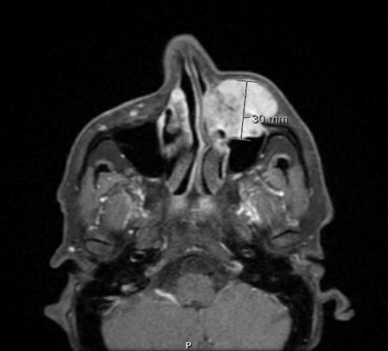
Initial magnetic resonance imaging of the face and neck showed a left maxillary mass, measuring 23 mm × 29 mm × 30 mm in the anteroposterior, mediolateral, and craniocaudal dimensions, respectively (Figure 1). Postcontrast sequences showed intense homogeneous enhancement and invasion of the left maxillary sinus through the anterior bony wall, resulting in bone destruction, and subsequent obstruction of the left nasal cavity. PET-CT scan after neoadjuvant radiotherapy, surgical excision, and three cycles of gemictabine chemotherapy showed a mild fludeoxyglucose avid, ill-defined, semisolid nodule in the right upper lobe, measuring 1.2 cm, and focal moderate to intense activity at the lateral aspect of the left clavicle. The patient was administered six cycles of gemcitabine and docetaxel chemotherapy. The latest follow-up PET-CT scan in May 2023 showed focal intense FDG uptake in the ascending colon and transverse colon, as well as liver metastasis. He was diagnosed with a pleomorphic leiomyosarcoma of the left maxilla with metastasis to the colon.

Figure 1
Figure 1 Magnetic resonance imaging of the face and neck showing a left maxillary mass.
FINAL DIAGNOSIS

Pleomorphic leiomyosarcoma of the left maxilla with metastasis to the colon.

TREATMENT

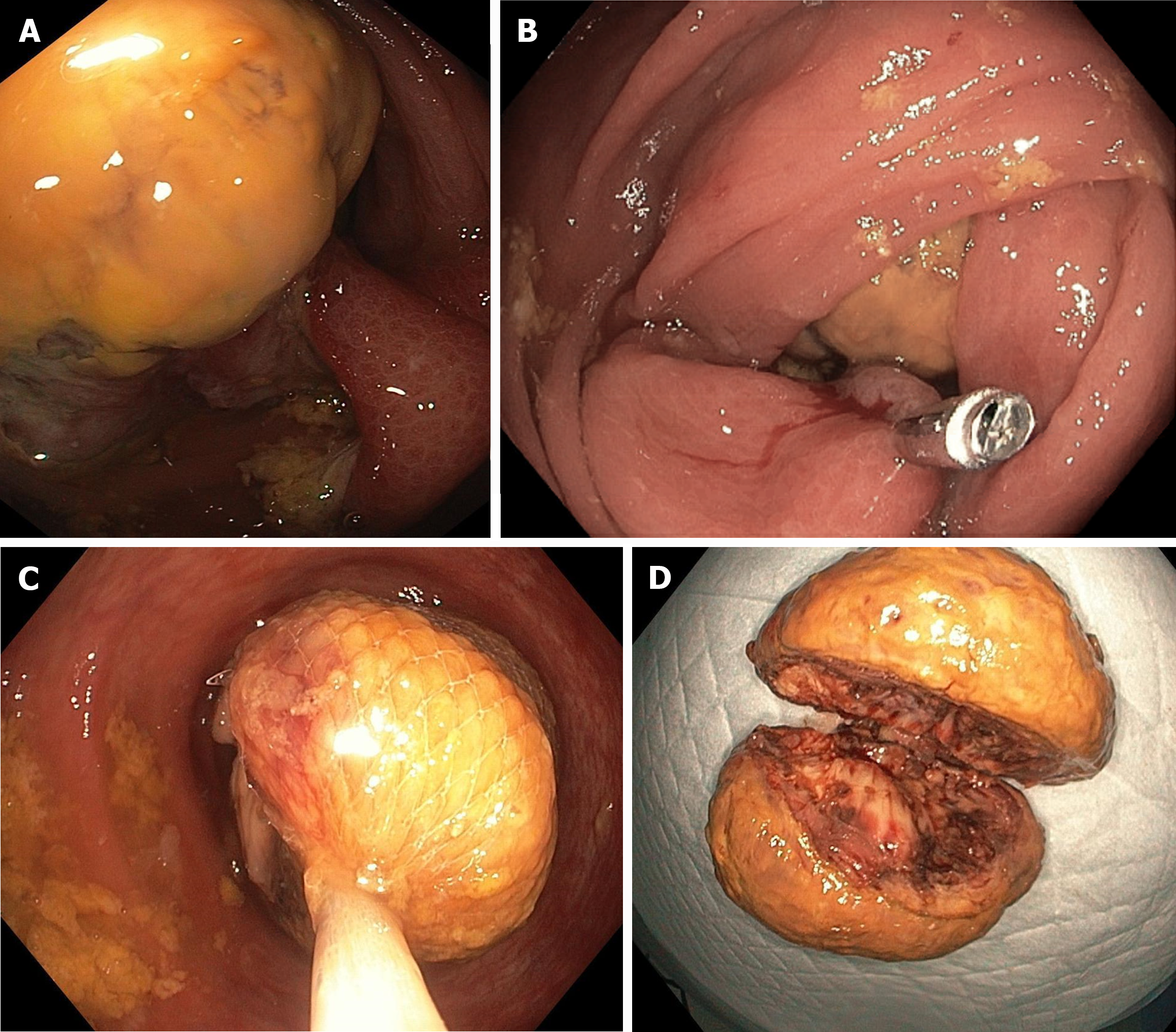
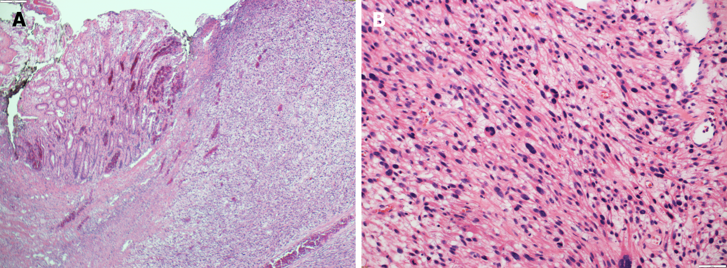
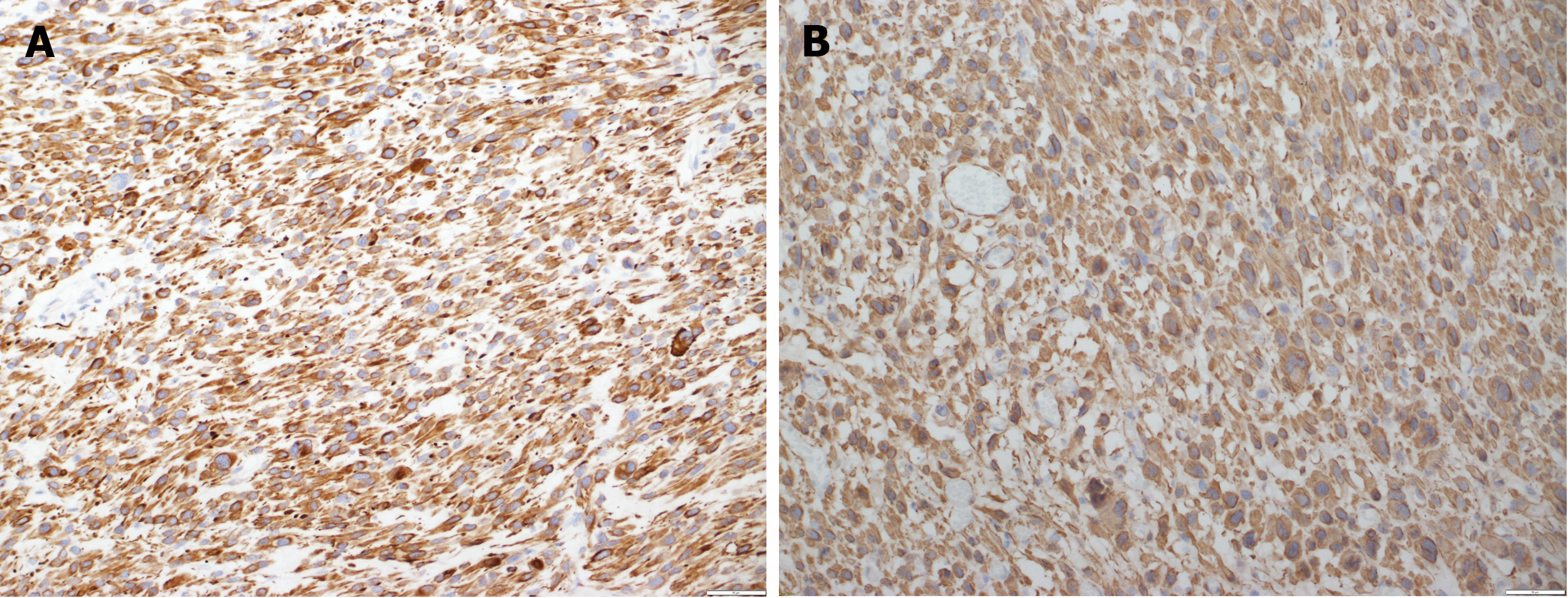
The patient was administered neoadjuvant radiation, followed by a left anterior maxillectomy. Pathologic examination confirmed that the tumor was a pleomorphic leiomyosarcoma of infraorbital soft tissue. Intraneural invasion was observed, but there was no evidence of lymphovascular invasion. The patient completed three cycles of doxorubicin chemotherapy. A follow-up PET-CT scan showed a mild FDG-avid, ill-defined semisolid nodule in the right upper lobe, measuring 1.2 cm, and focal moderate to intense activity at the lateral aspect of the left clavicle. Six cycles of gemcitabine and docetaxel chemotherapy were administered. The latest follow-up PET-CT scan in May 2023 showed focal intense FDG uptake in the ascending colon and transverse colon, as well as liver metastasis. The patient denied any pertinent symptoms, such as abdominal pain, alternating bowel habits, and PR bleeding. Colonoscopy showed a sizeable pedunculated polyp with a thick stalk in the proximal ascending colon, partially obstructing the lumen on the way to the cecum (Figure 2A). Endoscopic examination showed that the polyp was about 5 cm in size. Although polypectomy using an EndoLoop 30 mm was attempted, it was unsuccessful due to the large size of the polyp. Therefore, a hemoclip measuring 20 mm was applied to the stalk (Figure 2B). The polyp was removed by hot snare polypectomy, with two rounds required owing to the large size and thickness of the polyp. The first half was retrieved using a Roth net (Figure 2C). The second half was initially retrieved using a Roth net, with a scope and suctioning used when it reached the rectum. Direct examination showed that the polyp was 5.0 cm × 3.5 cm × 3.0 cm in size (Figure 2D). Pathological examination revealed a pleomorphic spindle cell malignancy with a high mitotic rate (Figure 3). Immunohistochemical examination showed that the tumor cells were positive for desmin and smooth muscle actin (Figure 4), but negative for markers of gastrointestinal stromal tumor (GIST), such as DOG-1 and CD 117.

Figure 2
Figure 2 Colonoscopy. A: Ascending colon polyp; B: Hemoclip applied to ascending colon polyp; C: Roth net covering ascending colon polyp; D: Retrieved ascending colon polyp.
Figure 3
Figure 3 Histopathological examination. A: Submucosal diffuse spindle cell neoplasm [hematoxylin and eosin (H&E) × 10]; B: Higher magnification of the spindle cell neoplasm showed high mitotic figures and prominent nuclear pleomorphism (H&E × 20).
Figure 4
Figure 4 Immunohistochemistry. A: The malignant cells are positive with desmin [immunohistochemistry (IHC) × 20]; B: The malignant cells are positive with SMA (IHC × 20).
OUTCOME AND FOLLOW-UP

The patient was followed-up in the clinic afterward. He did not report any gastrointestinal-related symptoms, but mostly complained of left clavicular and shoulder pain. He was treated with six cycles of dacarbazine chemotherapy, but the most recent CT scan revealed disease progression, as evidenced by increases in sizes of pulmonary nodules and liver lesions. He was then shifted to pazopanib.

DISCUSSION

Pleomorphic leiomyosarcoma can involve the retroperitoneum; the gastrointestinal tract, including the liver and pancreas; the adrenal glands; the mesenteric vessels; the skin; the heart; and the intrascrotum area[7-16]. Clinical manifestations have included abdominal discomfort, lower gastrointestinal bleeding, cardiac tamponade, back pain, and enlargement of the testicles or scrotum[7-15]. Oral cavity leiomyosarcomas are rare, with about eighty cases described in the literature[17]. More than half of these tumors involved the mandible[18]. Unlike leiomyosarcomas of other organs, intraoral leiomyosarcomas exhibit nodal metastasis, particularly in the cervical lymph nodes[19]. They also metastasize to the lungs and liver[19]. The present patient had metastases not only to the lungs and liver but also to the colon. To our knowledge, there have been no reports of other patients with pleomorphic leiomyosarcoma of the maxilla who showed metastasis to the colon. To date, only nine patents with pleomorphic leiomyosarcoma involving the mesocolon have been described[20], with the most common sites of involvement being the sigmoid and descending mesocolons. Mass, followed by pain, were the most common complaints. One case study described a patient who presented with lower GI bleeding secondary to mesenteric pleomorphic leiomyosarcoma that was inseparable from the descending colon[8]. The present patient presented with luminal involvement of the ascending colon with pleomorphic leiomyosarcoma, as evidenced by imaging and histopathologic examination of the polyp. Pleomorphic leiomyosarcomas are diagnosed through histological and immunohistochemical examination[7]. Typical histological features include smooth muscle cells, neoplastic spindle cells, and distinctly atypical pleomorphic cells[5,6]. Immunohistochemical staining has shown that most cells are positive for smooth muscle actin (90%-95%) and desmin (70%-90%)[21], with some being positive for caldesmon[5]. In contrast, pleomorphic leiomyosarcomas are negative for the GIST markers c-kit, CD117 and CD34, allowing differentiation of these two tumor types[6,8,22]. Although surgical resection with complete resection margins is the mainstay of management of pleomorphic leiomyosarcomas[8], local recurrence has been observed in 44%-85% of patients[23,24]. These findings point to the need for a comprehensive treatment plan, including chemotherapy and radiation. Neoadjuvant chemotherapy can reduce tumor size before surgical resection[25]. Although several randomized clinical trials have shown that adjuvant chemotherapy can minimize metastasis in patients with pleomorphic leiomyosarcomas, but further research is required to assess its survival benefit[26]. Radiotherapy is effective in treating pleomorphic leiomyosarcomas of the trunk and extremities[27]. Despite treatment, however, recurrence and metastasis are still common, with Metastatic pleomorphic leiomyosarcoma being associated with poor survival[8].

CONCLUSION

In summary, pleomorphic leiomyosarcoma is a rare and aggressive form of leiomyosarcoma that can affect any organ. Although few such patients have been described to date, their prognosis remains poor. Further studies are needed to understand this malignancy and explore newer treatment options.

Footnotes

Provenance and peer review: Unsolicited article; Externally peer reviewed.

Peer-review model: Single blind

Specialty type: Gastroenterology and hepatology

Country of origin: Saudi Arabia

Peer-review report’s classification

Scientific Quality: Grade C

Novelty: Grade C

Creativity or Innovation: Grade C

Scientific Significance: Grade C

P-Reviewer: Pattarajierapan S, Thailand S-Editor: Chen YL L-Editor: A P-Editor: Cai YX

References
1.  Serrano C, George S. Leiomyosarcoma. Hematol Oncol Clin North Am. 2013;27:957-974.  [PubMed]  [DOI]  [Cited in This Article: ]  [Cited by in Crossref: 84]  [Cited by in F6Publishing: 86]  [Article Influence: 7.8]  [Reference Citation Analysis (0)]
2.  Penel N, Coindre JM, Giraud A, Terrier P, Ranchere-Vince D, Collin F, Guellec SLE, Bazille C, Lae M, de Pinieux G, Ray-Coquard IL, Bonvalot S, Cesne ALE, Robin YM, Stoeckle E, Toulmonde M, Blay JY. Presentation and outcome of frequent and rare sarcoma histologic subtypes: A study of 10,262 patients with localized visceral/soft tissue sarcoma managed in reference centers. Cancer. 2018;124:1179-1187.  [PubMed]  [DOI]  [Cited in This Article: ]  [Cited by in Crossref: 26]  [Cited by in F6Publishing: 42]  [Article Influence: 6.0]  [Reference Citation Analysis (0)]
3.  Otabe M, Abe T, Sumi Y, Yonehara S, Noriyuki T, Nakahara M. Abnormal growth of a pleomorphic leiomyosarcoma originating from the mesenteric vein associated with poor outcome after curative-intent resection: a case report. Surg Case Rep. 2022;8:147.  [PubMed]  [DOI]  [Cited in This Article: ]  [Reference Citation Analysis (0)]
4.  Sandhu SV, Sodhi SP, Rai S, Bansal H. Primary leiomyosarcoma of the maxilla: An investigative loom-report of a challenging case and review of literature. J Oral Maxillofac Pathol. 2014;18:453-459.  [PubMed]  [DOI]  [Cited in This Article: ]  [Cited by in Crossref: 5]  [Cited by in F6Publishing: 5]  [Article Influence: 0.6]  [Reference Citation Analysis (0)]
5.  Oda Y, Miyajima K, Kawaguchi K, Tamiya S, Oshiro Y, Hachitanda Y, Oya M, Iwamoto Y, Tsuneyoshi M. Pleomorphic leiomyosarcoma: clinicopathologic and immunohistochemical study with special emphasis on its distinction from ordinary leiomyosarcoma and malignant fibrous histiocytoma. Am J Surg Pathol. 2001;25:1030-1038.  [PubMed]  [DOI]  [Cited in This Article: ]  [Cited by in Crossref: 107]  [Cited by in F6Publishing: 86]  [Article Influence: 3.7]  [Reference Citation Analysis (0)]
6.  Chen E, O'Connell F, Fletcher CD. Dedifferentiated leiomyosarcoma: clinicopathological analysis of 18 cases. Histopathology. 2011;59:1135-1143.  [PubMed]  [DOI]  [Cited in This Article: ]  [Cited by in Crossref: 35]  [Cited by in F6Publishing: 40]  [Article Influence: 3.3]  [Reference Citation Analysis (0)]
7.  Wang Y, Teng Y, Na S, Yuan Y. Pleomorphic Leiomyosarcoma of the Adrenal Gland in a Young Woman: A Case Report and Review of the Literature. Onco Targets Ther. 2020;13:4705-4713.  [PubMed]  [DOI]  [Cited in This Article: ]  [Cited by in Crossref: 3]  [Cited by in F6Publishing: 4]  [Article Influence: 1.0]  [Reference Citation Analysis (0)]
8.  Singh V, Aggarwal L, Hazrah P, Ali S, Kumar A, Mohta A. Pleomorphic leiomyosarcoma of colonic mesentery: an unusual cause of lower GI bleed - case report and literature review. Int Surg J. 2021;8:754.  [PubMed]  [DOI]  [Cited in This Article: ]
9.  Sagara K, Takayoshi K, Kusumoto E, Uchino K, Matsumura T, Kusaba H, Momosaki S, Ikejiri K, Baba E. Favorable control of rapidly progressive retroperitoneal pleomorphic leiomyosarcoma with multimodality therapy: a case report. BMC Res Notes. 2014;7:377.  [PubMed]  [DOI]  [Cited in This Article: ]  [Cited by in Crossref: 2]  [Cited by in F6Publishing: 2]  [Article Influence: 0.2]  [Reference Citation Analysis (0)]
10.  Fernández-Ferreira R, Soberanis-Piña PD, Conde-Flores E, Arroyave-Ramírez AM, Mackinney-Novelo I, Motola-Kuba D, Ruiz-Morales JM, Alvarado-Luna G, Chable-Montero F. Acute Cardiac Tamponade Secondary to Cardiac Vascular-Type Pleomorphic Leiomyosarcoma: Case Report. Case Rep Oncol. 2021;14:1860-1867.  [PubMed]  [DOI]  [Cited in This Article: ]  [Cited by in Crossref: 1]  [Cited by in F6Publishing: 1]  [Article Influence: 0.3]  [Reference Citation Analysis (0)]
11.  Smith CT, Grider DJ. Pleomorphic Leiomyosarcoma Presenting as a Upper Back Epidermal Inclusion Cyst. Am J Dermatopathol. 2021;43:446-449.  [PubMed]  [DOI]  [Cited in This Article: ]  [Reference Citation Analysis (0)]
12.  Popov H, Petkova L, Stoyanov GS. Lingual Leiomyosarcoma: A Histopathological Case Report. Cureus. 2022;14:e32717.  [PubMed]  [DOI]  [Cited in This Article: ]  [Reference Citation Analysis (0)]
13.  Damle RP, Suryawanshi KH, Dravid NV, Newadkar DV. Primary intratesticular pleomorphic leiomyosarcoma: a rare case report. Indian J Pathol Microbiol. 2013;56:425-427.  [PubMed]  [DOI]  [Cited in This Article: ]  [Cited by in Crossref: 2]  [Cited by in F6Publishing: 3]  [Article Influence: 0.3]  [Reference Citation Analysis (0)]
14.  Teshima T, Takahashi S, Nagamoto S, Miyazaki H, Nakagawa T, Fujimura T, Fukuhara H, Kume H, Homma Y. Intrascrotal dedifferentiated leiomyosarcoma originating from dartos muscle. Case Rep Urol. 2014;2014:841929.  [PubMed]  [DOI]  [Cited in This Article: ]  [Reference Citation Analysis (0)]
15.  Nesi G, Pantalone D, Ragionieri I, Amorosi A. Primary leiomyosarcoma of the pancreas: a case report and review of literature. Arch Pathol Lab Med. 2001;125:152-155.  [PubMed]  [DOI]  [Cited in This Article: ]  [Cited by in Crossref: 25]  [Cited by in F6Publishing: 24]  [Article Influence: 1.0]  [Reference Citation Analysis (0)]
16.  Enzinger FM, Weiss SW, Goldblum JR.   Leiomyosarcoma. Soft tissue tumours. 5th ed. St Louis: Mosby, 2008: 546-559.  [PubMed]  [DOI]  [Cited in This Article: ]
17.  Moghadam SA, Khodayari A, Mokhtari S. Primary leiomyosarcoma of the mandible. J Oral Maxillofac Pathol. 2014;18:308-311.  [PubMed]  [DOI]  [Cited in This Article: ]  [Cited by in Crossref: 2]  [Cited by in F6Publishing: 2]  [Article Influence: 0.2]  [Reference Citation Analysis (0)]
18.  Mendonça EF, Martins da Silva C, Meneghini AJ, Silva GB, Filho JA, Batista AC. Low-grade gingival leiomyosarcoma in a child. J Dent Child (Chic). 2008;75:301-305.  [PubMed]  [DOI]  [Cited in This Article: ]
19.  Dry SM, Jorgensen JL, Fletcher CD. Leiomyosarcomas of the oral cavity: an unusual topographic subset easily mistaken for nonmesenchymal tumours. Histopathology. 2000;36:210-220.  [PubMed]  [DOI]  [Cited in This Article: ]  [Cited by in Crossref: 50]  [Cited by in F6Publishing: 52]  [Article Influence: 2.2]  [Reference Citation Analysis (0)]
20.  Kajamohideen S, Venkitaraman B, Shivkumaran SM, Premkumar P. Primary pleomorphic leiomyosarcoma of descending mesocolon. Formos J Surg. 2021;54:196-199.  [PubMed]  [DOI]  [Cited in This Article: ]  [Reference Citation Analysis (0)]
21.  Deshmukh SD, Babanagare SV, Anand M, Pande DP, Yavalkar P. Primary adrenal leiomyosarcoma: a case report with immunohistochemical study and review of literature. J Cancer Res Ther. 2013;9:114-116.  [PubMed]  [DOI]  [Cited in This Article: ]  [Cited by in Crossref: 18]  [Cited by in F6Publishing: 18]  [Article Influence: 1.6]  [Reference Citation Analysis (0)]
22.  Hirota S, Isozaki K, Moriyama Y, Hashimoto K, Nishida T, Ishiguro S, Kawano K, Hanada M, Kurata A, Takeda M, Muhammad Tunio G, Matsuzawa Y, Kanakura Y, Shinomura Y, Kitamura Y. Gain-of-function mutations of c-kit in human gastrointestinal stromal tumors. Science. 1998;279:577-580.  [PubMed]  [DOI]  [Cited in This Article: ]  [Cited by in Crossref: 3215]  [Cited by in F6Publishing: 3056]  [Article Influence: 117.5]  [Reference Citation Analysis (0)]
23.  Pervaiz N, Colterjohn N, Farrokhyar F, Tozer R, Figueredo A, Ghert M. A systematic meta-analysis of randomized controlled trials of adjuvant chemotherapy for localized resectable soft-tissue sarcoma. Cancer. 2008;113:573-581.  [PubMed]  [DOI]  [Cited in This Article: ]  [Cited by in Crossref: 640]  [Cited by in F6Publishing: 635]  [Article Influence: 39.7]  [Reference Citation Analysis (0)]
24.  Weitz J, Klimstra DS, Cymes K, Jarnagin WR, D'Angelica M, La Quaglia MP, Fong Y, Brennan MF, Blumgart LH, Dematteo RP. Management of primary liver sarcomas. Cancer. 2007;109:1391-1396.  [PubMed]  [DOI]  [Cited in This Article: ]  [Cited by in Crossref: 133]  [Cited by in F6Publishing: 121]  [Article Influence: 7.1]  [Reference Citation Analysis (0)]
25.  Matthaei H, Krieg A, Schmelzle M, Boelke E, Poremba C, Rogiers X, Knoefel WT, Peiper M. Long-term survival after surgery for primary hepatic sarcoma in adults. Arch Surg. 2009;144:339-44; discussion 344.  [PubMed]  [DOI]  [Cited in This Article: ]  [Cited by in Crossref: 51]  [Cited by in F6Publishing: 45]  [Article Influence: 3.0]  [Reference Citation Analysis (0)]
26.  Bathan AJ, Constantinidou A, Pollack SM, Jones RL. Diagnosis, prognosis, and management of leiomyosarcoma: recognition of anatomic variants. Curr Opin Oncol. 2013;25:384-389.  [PubMed]  [DOI]  [Cited in This Article: ]  [Cited by in Crossref: 31]  [Cited by in F6Publishing: 34]  [Article Influence: 3.1]  [Reference Citation Analysis (0)]
27.  Gupta S, Husain N, Kumar A, Sunder S. Adult primary pleomorphic leiomyosarcoma of forearm with axillary lymph node metastasis: a case report and literature review. J Cancer Res Ther. 2012;8:448-450.  [PubMed]  [DOI]  [Cited in This Article: ]  [Cited by in Crossref: 2]  [Cited by in F6Publishing: 2]  [Article Influence: 0.2]  [Reference Citation Analysis (0)]