Copyright
©The Author(s) 2018.
World J Gastrointest Endosc. Nov 16, 2018; 10(11): 326-339
Published online Nov 16, 2018. doi: 10.4253/wjge.v10.i11.326
Published online Nov 16, 2018. doi: 10.4253/wjge.v10.i11.326
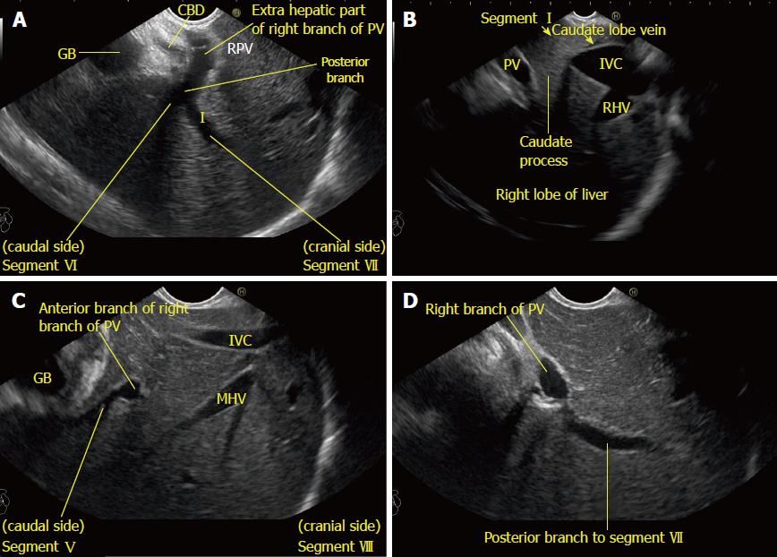
Figure 7 Imaging from station 1 showing the right portal vein and its branches in relation to liver segments.
A: Imaging of the intrahepatic/extrahepatic part of the right branch of the portal vein is seen along with the right lobe of the liver and the hyperechoic diaphragm; B: The inferior vena cava and caudate process provide a good window of imaging for the right lobe of the liver (The caudate lobe is connected with the right lobe of liver through the caudate process). In this location, presence of the inferior vena cava may also provide a satisfactory window of imaging; C: A view of the divisions of the right branch of the portal vein is possible through the caudate process. In this case, the anterior branch is supplying segments V and VIII of the liver; D: The division of segment VI and VII branches is visualized. The upper part of the posterior branch goes towards segment VII. CBD: Common bile duct; GB: Gallbladder; PV: Portal vein; RHV: Right hepatic vein; MHV: Middle hepatic vein; IVC: Inferior vena cava.
- Citation: Sharma M, Somani P, Rameshbabu CS, Sunkara T, Rai P. Stepwise evaluation of liver sectors and liver segments by endoscopic ultrasound. World J Gastrointest Endosc 2018; 10(11): 326-339
- URL: https://www.wjgnet.com/1948-5190/full/v10/i11/326.htm
- DOI: https://dx.doi.org/10.4253/wjge.v10.i11.326


