Copyright
©The Author(s) 2018.
World J Gastrointest Endosc. Nov 16, 2018; 10(11): 326-339
Published online Nov 16, 2018. doi: 10.4253/wjge.v10.i11.326
Published online Nov 16, 2018. doi: 10.4253/wjge.v10.i11.326
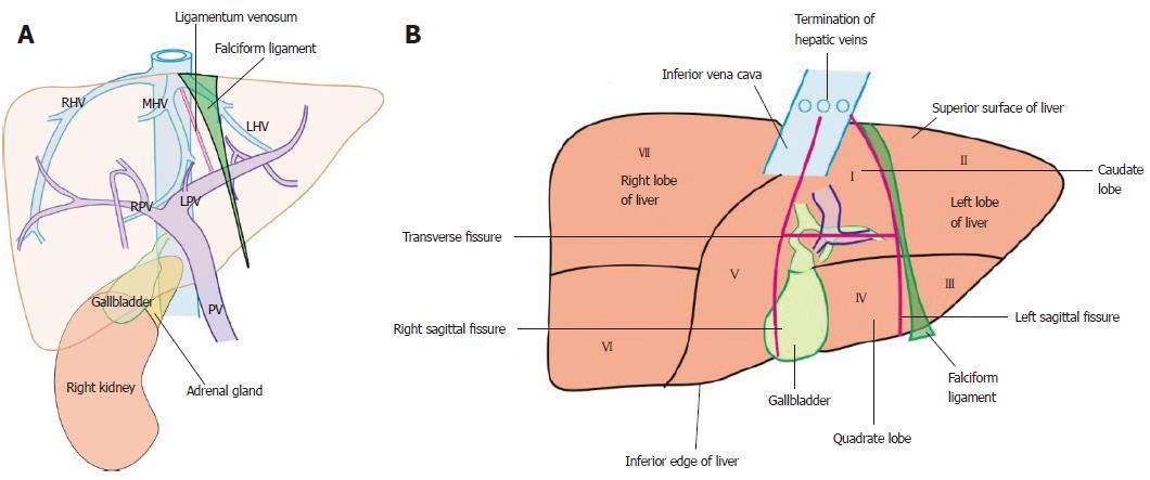
Figure 2 The home bases of imaging and visceral surface of liver.
A: The endoscopic ultrasonography home bases of imaging for liver segments include the inferior vena cava (IVC) during its course behind the liver up to the right atrium, three hepatic veins in portal fissures and their joining point into the IVC, the portal vein and its branches in transverse fissure, the right kidney, the ligamentum teres, the ligamentum venosum, and the gallbladder; B: The caudate lobe lies between the stomach and IVC. The hepatic veins join the IVC. The ligamentum venosum and ligamentum teres are attached to the upper and lower border of the left branch of the portal at the angulation between the transverse and umbilical part. The visceral surface of the liver comes in contact with all segments except segment VIII. PV: Portal vein; LHV: Left hepatic vein; MHV: Middle hepatic vein; RHV: Right hepatic vein; LPV: Left branch of the portal vein; RPV: Right branch of the portal vein.
- Citation: Sharma M, Somani P, Rameshbabu CS, Sunkara T, Rai P. Stepwise evaluation of liver sectors and liver segments by endoscopic ultrasound. World J Gastrointest Endosc 2018; 10(11): 326-339
- URL: https://www.wjgnet.com/1948-5190/full/v10/i11/326.htm
- DOI: https://dx.doi.org/10.4253/wjge.v10.i11.326


