Published online Nov 16, 2018. doi: 10.4253/wjge.v10.i11.326
Peer-review started: May 25, 2018
First decision: June 14, 2018
Revised: August 16, 2018
Accepted: October 17, 2018
Article in press: October 17, 2018
Published online: November 16, 2018
Processing time: 174 Days and 23.1 Hours
The liver has eight segments, which are referred to by numbers or by names. The numbering of the segments is done in a counterclockwise manner with the liver being viewed from the inferior surface, starting from Segment I (the caudate lobe). Standard anatomical description of the liver segments is available by computed tomographic scan and ultrasonography. Endoscopic ultrasound (EUS) has been used for a detailed imaging of many intra-abdominal organs and for the assessment of intra-abdominal vasculature. A stepwise evaluation of the liver segments by EUS has not been described. In this article, we have described a stepwise evaluation of the liver segments by EUS. This information can be useful for planning successful radical surgeries, preparing for biopsy, portal vein embolization, transjugular intrahepatic portosystemic shunt, tumour resection or partial hepatectomy, and for planning EUS guided diagnostic and therapeutic procedures.
Core tip: Standard anatomical description of the liver segments is available by computed tomographic scan and ultrasonography. A stepwise evaluation of the liver segments by endoscopic ultrasound (EUS) has not been described. In this article, we have described a stepwise evaluation of the liver segments by EUS. This information can be useful for planning successful radical surgeries, preparing for biopsy, portal vein embolization, transjugular intrahepatic portosystemic shunt, tumour resection or partial hepatectomy, and for planning EUS guided diagnostic and therapeutic procedures.
- Citation: Sharma M, Somani P, Rameshbabu CS, Sunkara T, Rai P. Stepwise evaluation of liver sectors and liver segments by endoscopic ultrasound. World J Gastrointest Endosc 2018; 10(11): 326-339
- URL: https://www.wjgnet.com/1948-5190/full/v10/i11/326.htm
- DOI: https://dx.doi.org/10.4253/wjge.v10.i11.326
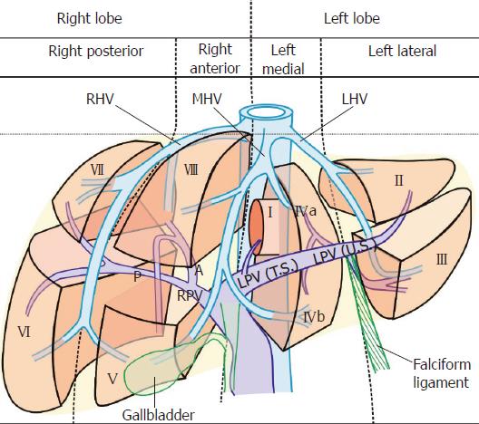
The French surgeon and anatomist Claude Couinaud described “two lobes” (right and left), “two hemilivers” (right and left), “four sectors,” and “eight segments” of the liver. The two lobes are separated by the falciform ligament, the two hemilivers are separated by the Cantlie’s line, the four sectors are separated by the planes of three hepatic veins (portal fissures or scissurae), and the eight segments are separated by an imaginary transverse plane passing by the portal vein bifurcation (Figure 1 and Table 1)[1]. Each of the segments is an independent segment and has its own vascular inflow, outflow, and biliary drainage. The segments are referred to by number or by names[2,3]. The numbering of the segments is done in a counterclockwise manner starting from Segment I (the caudate lobe). The segments II to IV belong to the left and segments V to VIII belong to the right hemiliver (Table 1)[2-4]. Standard anatomical description of the liver segments is available by computed tomographic (CT) scan[5]. A stepwise evaluation of the liver segments is also described by ultrasonography[6,7]. Endoscopic ultrasound (EUS) has been used for detailed imaging of many intra-abdominal organs and for the assessment of intra-abdominal vasculature[8-11]. A stepwise evaluation of the liver segments by EUS has not been described. In this article, we describe a stepwise evaluation of the liver segments by EUS.
| Four hepatic sectors | Eight hepatic segments |
| Segment I - the caudate lobe | |
| Left lateral sector | Segment II - posterosuperior |
| Segment III - anteroinferior | |
| Left medial sector | IVa - superior segment |
| IVb - inferior segment | |
| Right anterior sector | Segment V - inferior segment |
| Segment VIII - superior segment | |
| Right posterior sector | Segment VI - inferior segment |
| Segment VII - superior segment |
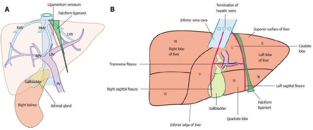
The home bases of liver imaging are given in the figure and the table (Figure 2A and Table 2). The main key of the imaging is following the hepatic vein tributaries and the portal vein branches. The three hepatic veins divide the liver into four vertical sectors: right anterior, right posterior, left medial and left lateral (Figure 1). The plane of portal vein bifurcation creates a transverse plane that crosses the vertical planes and divides the four sectors into eight segments (Figures 1 and 2A, and Table 1). The right hepatic vein lies in the right portal fissure and separates the right hemiliver into anterior and posterior sectors. The left hepatic vein lies in the left portal fissure and separates the left hemiliver into medial and lateral sectors. The middle hepatic vein lies in the main portal fissure and separates the anterior division of right liver from the medial division of the left liver. The main portal vein bifurcates into the right and the left branches, which travel in an imaginary transverse plane of the transverse fissure. The extrahepatic part of the left branch of the portal vein is known as the transverse part and the intrahepatic part of left branch of the portal vein is known as the umbilical part. The extrahepatic part of left branch of portal vein lies in the gastrohepatic ligament on its inferior surface. The umbilical part of portal vein is surrounded on all sides by the liver (Figure 1). The right portal vein divides into two branches after entering the liver. The anterior branch supplies the anterior sector of the right lobe, and the posterior branch supplies the posterior sector of the right lobe (Figures 1 and 2A, and Table 2). The gallbladder, the right kidney, the fissures on the under surface of liver, and the ligaments of liver are acting as additional home bases of imaging for liver segments.
| Home bases | Defining of segments |
| Hepatic veins | LHV separates II and III from IV (a and b) |
| MHV separates IVa from VIII and IVb from V | |
| RHV separates V from VI and VIII from VII | |
| Portal vein and its branches | Superior segments lie above and inferior segments lie below the transverse fissure |
| The LPV supplies segments II to IV | |
| The RPV supplies segments V to VIII | |
| IVC | IVC passes through the bare area of the liver and is related to superior segments in the upper part, segment I for most of its course, and with segment VI close to the lower most part above the right kidney |
| IVC suprahepatic part | A transverse plane defines the upper limit of superior segments (II, IV, VIII, and VII) |
| Ligamentum teres | Separates segment III from IVb |
| Ligamentum venosum | Separates I from IVa and II |
| Gallbladder | Neck lies close to segment V and fundus close to segment VI, from stomach IVb comes between the gallbladder and probe |
The inferior surface of the liver has an H-shaped fissure where the right and left limbs of the H are made by the right and left sagittal fissures, and the transverse limb is formed by the porta hepatis. The left sagittal fissure has upper and lower limbs, which are formed by the fissure for the ligamentum venosum and the fissure for the ligamentum teres. The upper part of the ligamentum venosum is attached to the inferior vena cava (IVC). The lower part of the ligamentum teres extends to the inferior surface of liver (Figure 2B and Table 2). The relationship of the caudate lobe is crucial in understanding the segments of liver. When seen from the stomach the caudate lobe lies between the stomach and the IVC. The hepatic veins join the IVC (Figure 2B).
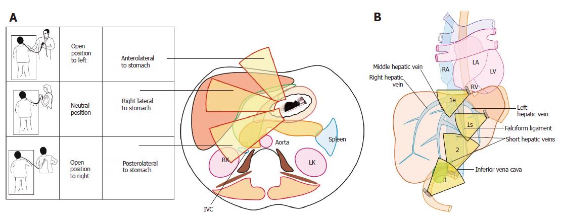
EUS can be done with the patient in a prone, left lateral, or supine position, and the descriptions in this article have been done with the patient in a prone position. The position of the operator can change with the movement of the body (or hand), which transfers the effect of rotation towards the tip of the scope when the scope is maintained in a straight position. The three positions are open position to left, neutral position, and an open position to right. In an open position to left, the operator faces the patient’s feet. In a neutral position, the operator faces the body of the patient. In an open position to right, the operator faces the head of the patient. A clockwise rotation from an open position to left in the stomach moves the imaging axis of the probe from a dorsal to a lateral and subsequently to a ventral position (Figure 3A). A reverse of this happens when the scope is rotated counterclockwise after seeing the right kidney.
The imaging is possible from three stations. Station 1: abdominal part of esophagus and stomach; station 2: duodenal bulb; and station 3: 2nd part of duodenum (Figure 3B and Table 3). Each station shows the segments in different anatomical relationships, and the differences in the segmental assessment of imaging are described in the table.
| Station | Comment | Segments visualized |
| Abdominal part of the esophagus and stomach | Probe in the esophagus lies close to the left lobe of the liver and in the stomach close to the visceral surface of the liver | Superior segments and caudate lobe are in direct contact with the lower end of esophagus All segments except segment VIII on the visceral surface of liver are in contact with the stomach |
| Duodenal bulb | Probe lies close to the hilum of the liver | Direct contact with segment I and right lobe liver segments Left side segments are seen through caudate lobe, caudate process, and IVC |
| Second part of duodenum. | Probe lies close to the hilum of the liver | Direct contact with segment I and right lobe liver segments Left side segments are seen through caudate lobe, caudate process, and IVC |
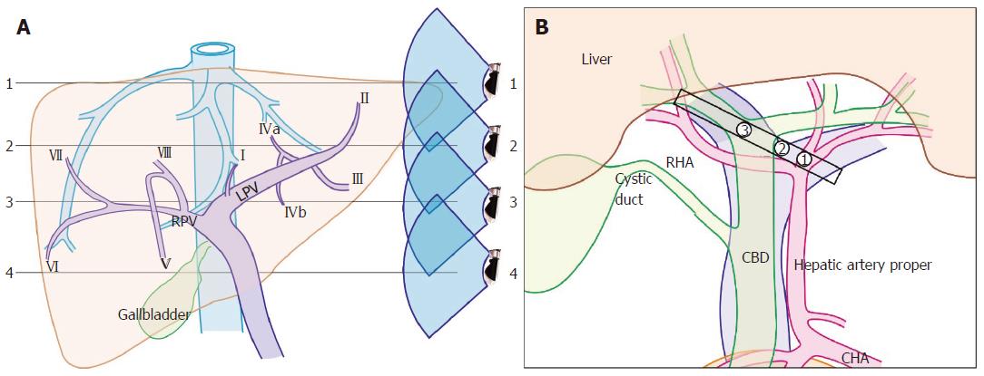
This station of imaging is the most convenient method of imaging and is discussed in detail. Generally, it requires a rotation of the scope in three positions (Figure 3A) and a shift of the scope in and out to the four different levels (Figure 4A). During this rotation the sectors come close to the probe in the following order left lateral, left medial, right anterior, and right posterior (Table 4).
| Clockwise rotation from an open position to left | Part of portal vein | Part of hepatic vein | Other home base structures | Main sector of liver visualized | Main segments of liver visualized | Figure number for segment |
| Open position to left | PII and PIII | LHV | Diaphragm and heart | Left medial closer to left lateral1 | II, III, and IV | 2A, 2B, 3A, 5A |
| Neutral position after approximately 60° to 75° clockwise rotation | Fisheye appearance of LPV | MHV | Left edge of transverse fissure attached to ligamentum venosum and ligamentum teres | Left medial closer to probe than right anterior | I and IV | 3A, 5B |
| Open position to right after further approximately 60° to 75° clockwise rotation | RPV dividing into anterior and posterior branches | RHV | Right edge of transverse fissure, IVC, gallbladder, and caudate process | Right anterior closer to probe than right posterior | V, VI, VII, and VIII | 3A, 7A 7C, 7D |
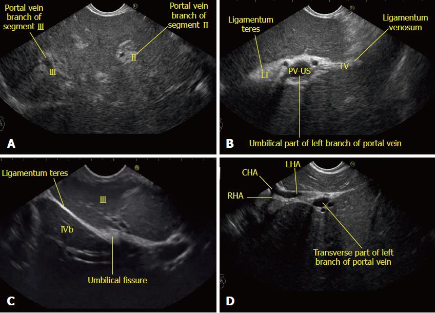
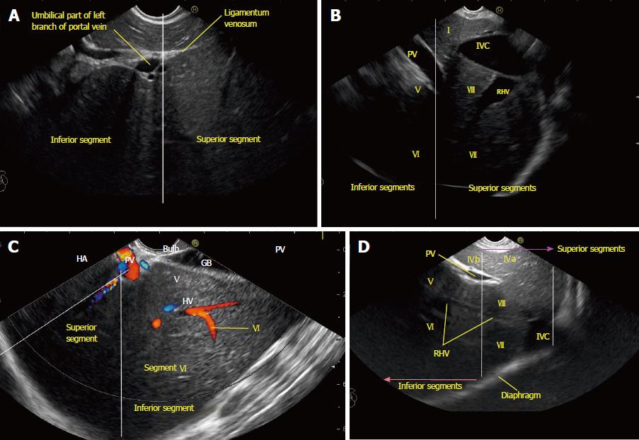
Imaging from open position to left: From the open position to left, the portal vein branches going to segment II and III are identified (Figure 5A). Imaging of the intrahepatic part of the left branch of the portal vein (umbilical part) within the umbilical fissure gives a fisheye appearance as it is completely surrounded on all sides by the liver parenchymal tissue (Figure 5B). The fisheye appearance indicates the left edge of the H of the transverse fissure from where the upper and lower limbs of the left sagittal fissure can be followed by slight in and out movement to trace the ligamentum venosum and ligamentum teres (Figures 5B and 5C). Further rotation traces the transverse fissure from the left edge of the H towards the right edge of the H and follows the umbilical part of the left branch of the portal vein towards the transverse part of the left branch. During this rotation from the left edge to the right edge, the appearance of the hepatic artery into two branches, the union of right and left branch of the portal vein and the division of the common hepatic duct into right and left branches appear one by one in the transverse fissure (Figures 4B and 5D).
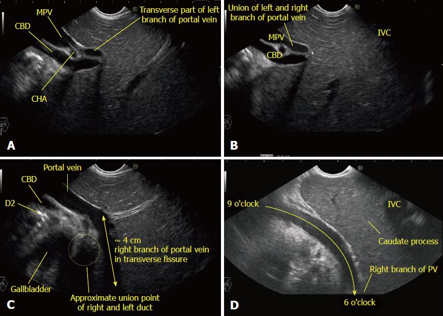
Imaging from the neutral position: Further rotation traces the course of the extrahepatic part of the left branch of the portal vein (transverse part) within the transverse fissure (Figure 6A). At this point the union of right and left branches of the portal vein is seen (Figure 6B). Near the union point the extrahepatic part of the right branch of the portal vein can be seen to join the transverse part of the portal vein from a direction coming from 6 o’clock within the transverse fissure. The right branch of the portal vein is often seen traveling close to the neck of gallbladder. Near the right edge of the transverse fissure, the union of right and left ducts is seen in front of the right branch of the portal vein (Figure 6C). The imaging of the right branch of the portal vein is easy through the caudate process of the liver in the area above the transverse fissure and below the IVC (Figure 6D).
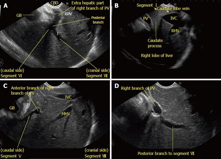
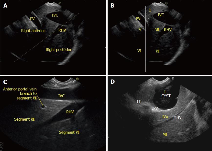
Imaging from an open position to right: Further rotation with slight upward angulation traces the extrahepatic part of the right branch of the portal vein towards the intrahepatic part. Imaging of the intrahepatic part of the right branch of the portal vein usually requires a decrease in frequency to increase penetration depth and an increase in the depth of the imaging to include the entire right lobe of the liver along with the hyperechoic diaphragm, which is seen beyond the convex upper surface of the liver (Figure 7A). Imaging of the right part of liver is easy due to the window made by the IVC and the caudate process (Figure 7B). The right branch of the portal vein can be followed to the division into an anterior and a posterior branch. Usually, with a linear scope the vision of the anterior branch is best seen when the middle hepatic vein is seen on the far side of the screen (Figure 7C). A relatively more posterior view of the course of posterior division of the right portal vein branches is possible through the caudate process between the porta hepatis and the IVC. It is usually possible to see the segmental branches to segment VI and VII (Figure 7D).
Neutral position: Imaging from duodenal bulb shows a view of the liver hilum (transverse fissure) where the portal vein is easily identified, with right and left angulation, up and down movements, and in and out adjustments.
Open position to left: The portal vein bifurcation defines the line between superior and inferior segments. A counterclockwise rotation traces the transverse fissure towards the gallbladder and the right edge of the transverse fissure where the right lobe segments are seen.
Open position to right: A clockwise rotation after tracing the portal vein bifurcation traces the transverse fissure towards the left edge of the transverse fissure where the left lobe segments are seen. The lower border of the caudate lobe lies above the bifurcation and above the transverse part of the left branch of the portal vein.
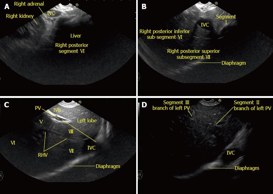
An open position to right places the scope in a parallel axis with the superior mesenteric vein from the descending duodenum. From this position a counterclockwise rotation traces the course of the IVC from a 10 o’clock position to a 4 o’clock position and sequentially brings the hilum of the right kidney, the right lobe of liver, the hilum of the liver and gallbladder, and lastly the left lobe of the liver into view (Figure 8). In this process the segments belonging to the posterior surface and bare area of the liver are easily followed close to the IVC. The first segment close to the lower most part of the IVC just above the adrenal gland and right kidney belongs to segment VI (Figure 8A). Once the gallbladder is seen segment V lies close to the upper surface of the gallbladder. Near the hilum of the liver segment I is identified between the portal vein and the IVC. On maximum counterclockwise rotation the probe lies close to the upper most part of the IVC near the joining of the hepatic veins where the superior segments (VII, VIII, IVa, and II) are visualized.
The imaging of segments is done in eight steps: (1) identify lobes; (2) identify sectors (right and left portal fissures); (3) identify the plane of superior and inferior segments (transverse fissure/liver hilum); (4) identify left sagittal fissure and the liver ligaments; (5) identify caudate lobe (posterior to transverse fissure); (6) identify left lobe segments; (7) identify quadrate lobe (anterior to transverse fissure); and (8) identify right lobe segments.
Applied anatomy: The Cantlie’s line passes through the middle hepatic vein (main portal fissure) superiorly. It also corresponds to a vertical plane passing diagonally from the middle of the gallbladder fossa anteriorly and inferiorly to the left side of the IVC posteriorly (Figure 2B).
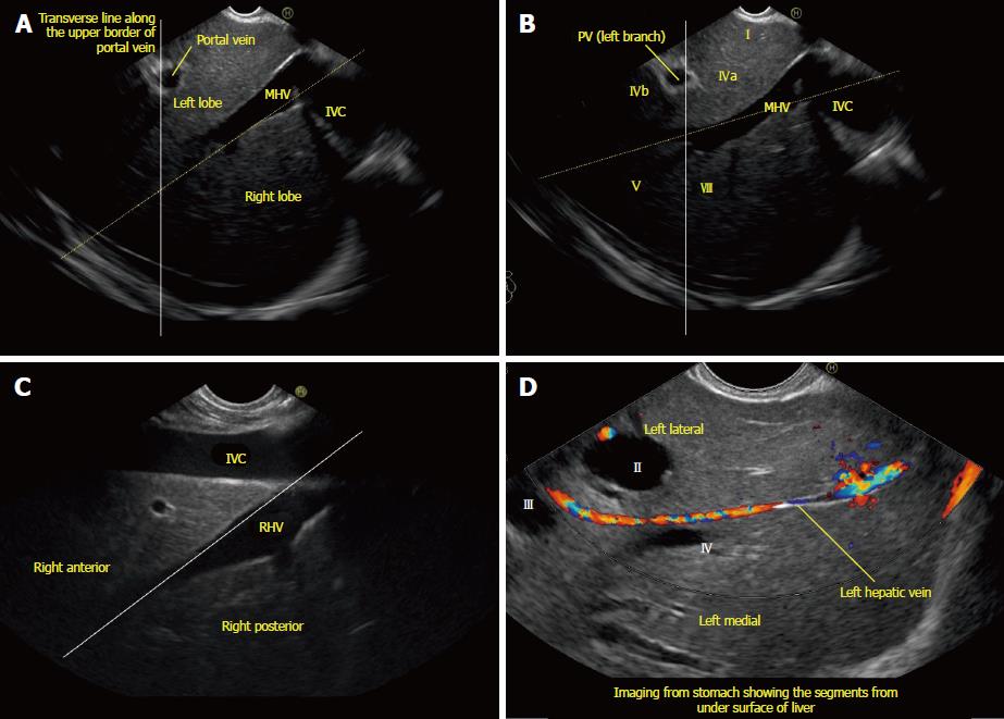
Technique of examination: Identify the IVC. The middle hepatic vein is identified merging into IVC (Figures 9A and 9B).
Applied anatomy: Each right and left hemiliver is subdivided by the left and right hepatic veins lying in the left and right portal fissures. The course of the left hepatic vein divides the left lobe into the medial and lateral sectors. The course of the right hepatic vein divides the right lobe into the anterior and posterior sectors.
Technique of examination: Identify the merger of the left and right hepatic veins into the IVC (Figures 9C and 9D).
Applied anatomy: The portal vein and its branches in the transverse fissure acts as a guide to divide the liver into superior (VII, VIII, IVa and II) and inferior segments (III, IVb V and VI). The applied anatomy of fissures and ligaments was already discussed in the home bases section. The joining of the three hepatic veins into the supra hepatic part of the IVC determines the uppermost margin of superior segments related to the IVC.
Technique of examination: The umbilical fissure is identified within the left lobe of the liver. Clockwise rotation along the umbilical fissure traces the transverse plane of the transverse fissure within which the left and right branches of the portal vein are seen (Figures 6C, 6D, and 10).
Applied anatomy: The applied anatomy of fissures and ligaments was already discussed in the home bases section.
Technique of examination: The umbilical fissure is identified within the left lobe of the liver. The ligaments are attached to the upper and lower part of the umbilical fissure (Figure 5C).
Applied anatomy: Anatomy texts describe the caudate lobe as a midline, vertically oriented hepatic lobe seen on the posterior aspect of the liver separating a portion of the right and left hepatic lobes in an H configuration. The horizontal bar of the H configuration represents the transverse fissure of the porta hepatis, which includes the horizontal portion of both portal veins. Above the bar is the caudate lobe of the liver and below the bar is the medial segment, or quadrate lobe, of the left lobe of the liver[12].
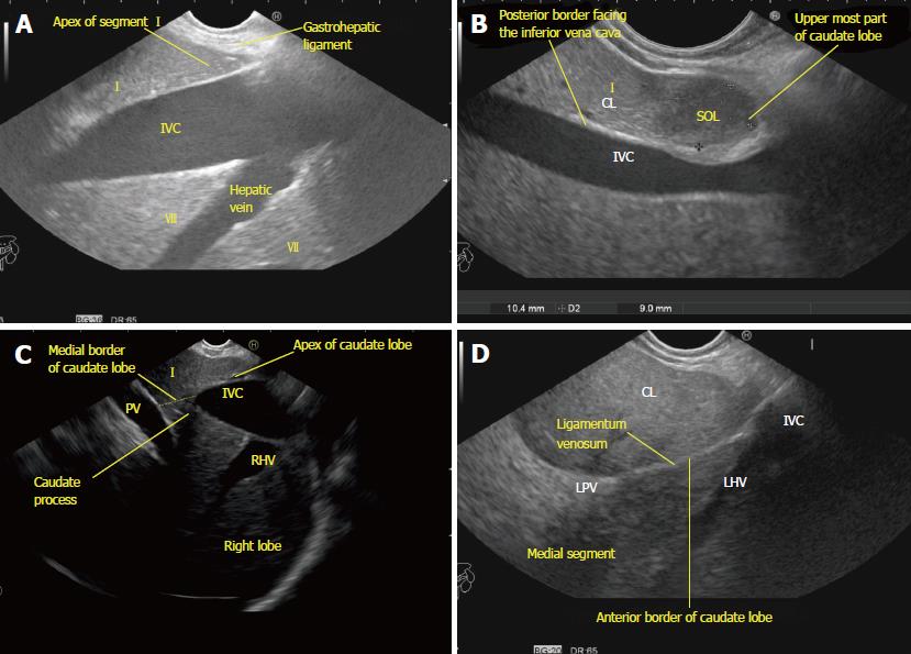
Technique of examination: The caudate lobe may be imagined as a midline wedge in a sagittal plane with its tip extending cephalad up to the insertion of the left and middle hepatic veins into the IVC and its base or posterior border facing the IVC (Figures 11A and 11B). The right (or medial border) of the pyramid is continuous with the parenchyma of the right lobe of the liver via the caudate process (Figure 11C). The anterior border of the caudate lobe is separated from the medial segment of the left lobe of the liver superiorly by the fissure for the ligamentum venosum and inferiorly by the left portal triad and portal bifurcation (Figure 11D). A clockwise rotation after identification of the left lobe of the liver from an open position to left will help in identification of the ligamentum venosum (Figure 11D). The upper border of the umbilical part of the left branch of the portal vein is attached to ligamentum venosum and serves as an accurate anatomic boundary of the lowermost limit and anterior margin of the caudate lobe (Figure 11D).
The caudal margin of the caudate lobe form the cephalad margin, or lintel, of the foramen of Winslow and projects into the lesser sac where it is related to the right crus of the diaphragm and inferior phrenic artery. The left (or lateral border) of the caudate lobe projects into the superior recess of the lesser peritoneal sac and is covered anteriorly by the gastrohepatic ligament (lesser omentum), which separates it from segments II and III anteriorly and segment VIII in the right lateral wall (Figure 12B). The vascular inflow and biliary drainage to the caudate lobe comes from both the right and left pedicles. The right side of the caudate, the caudate process, largely derives its portal venous supply from the right portal vein or the bifurcation of the main portal vein (Figure 12C). The left portion of the caudate derives its portal venous inflow from the left portal vein.
Caudate process a two-way window for imaging of the right and left lobe of the liver in EUS: The caudate lobe, caudate process, and the IVC act as a window of imaging from the left side to the right side of the liver and vice versa. While imaging from the abdominal part of the esophagus and stomach the window allows left to right visualization. While imaging from the duodenal bulb and the second part of the duodenum the window allows right to left visualization (Figures 11C, 11D, 12A, 12C, and 13).
Applied anatomy: The left lobe lies anterior to the esophagogastric junction and the fundus of stomach.
Technique of examination: The segments of the left lobe are best visualized with slight up tilting of the scope from the fundus of stomach. In this position, the left part of the heart is visualized through the diaphragm and the left hepatic vein is easily identified as the border between the left medial and two left lateral segments (segment III anterior-inferior and segment II posterior-superior). Further demarcation of segment II as the upper segment and segment III as the lower segment is possible by the plane passing by the portal vein bifurcation (Figures 14A and 14B). This plane is not usually visualized during imaging of the left lateral segments (II and III) in an open position to left where the fisheye appearance of the umbilical part is not visualized. A line can be extrapolated from the upper border of the fisheye appearance of the left branch of the portal vein for proper demarcation (Figure 14B). The visualization of the left medial segment is seen between the probe and the middle hepatic vein (Figure 14C). The presence of the middle hepatic vein and of the upper border of the left branch of the portal vein indicates the area where segment I communicates with segment IV (Figure 14D).
Identify segment IVa and IVb (quadrate lobe)
Applied anatomy: Segment IV is located between the plane passing by the middle hepatic vein on the right and the axis of the umbilical scissura on the left. This segment can be divided into an upper (IVa) and a lower (IVb) segment by a horizontal line passing through the umbilical portion of the left branch of the portal vein. The IVb segment (quadrate lobe) is demarcated on the visceral surface anteriorly by the inferior edge of the liver, on the right side by the gallbladder fossa, posteriorly by the porta hepatis, and on the medial side by the fissure for the ligamentum teres (Figures 14B and 14D).
Technique of examination: Segment IV is easily identified near the middle hepatic vein. The identification of the gallbladder, ligamentum teres, and umbilical fissure also helps in identification of the liver segments (Figure 14D).
Applied anatomy: The right liver consists of anterior and posterior segments, divided by the presence of the right hepatic vein in the right portal fissure (Figure 15A).
Technique of examination: A simple crossover of lines passing through the upper border of the left branch of the portal vein and along the axis of the right hepatic vein will divide the right hemiliver into four segments (Figure 15).
Table 5 summarizes the evaluation of different liver segments from different positions by EUS. Routine use of Couinaud’s liver segmentation may be important in planning successful radical surgery. This description will allow better preparation for biopsy, portal vein embolization, transjugular intrahepatic portosystemic shunt, tumour resection, or partial hepatectomy for transplantation. Such advance planning will reduce intra- and post-operative difficulties and complications. In this article, we have given a detailed description of EUS anatomy of the liver and liver segmentation. This information may be useful in planning EUS guided diagnostic and therapeutic procedures involving liver pathologies[13,14].
| Home base structure | Comment | Figure No. |
| Hepatic veins | The main left hepatic vein drains the two lateral segments of the left lobe (segments II and III) | 9D |
| The middle vein drains segment IV and the anterior sector of the right lobe (segments V and VIII) | 9B | |
| The right vein drains the remainder of the right lobe (segments VI and VII) and a variable portion of segments Vand VIII | 9C | |
| Hepatic vein tributaries | The higher tributary of LHV going towards the diaphragmatic and costal surface belongs to segment II | 9D |
| The higher tributary of MHV going towards the diaphragmatic and costal surface belongs to segment VIII | 9B, 14C, 14D | |
| The higher tributary of RHV going towards the diaphragmatic and costal surface belongs to segment VII | 15A, 15C | |
| The tributary of LHV going towards the liver hilum drains segment IVa | 14B | |
| The tributary of MHV going towards the liver hilum drains segment IVb | 14D | |
| The tributaries of RHV going towards the liver hilum drains segments Vand VIII | 9C | |
| Right and left branches of the portal vein | The right branch of the portal vein supplies the right liver (segments Vto VIII) | 7A, 7B, 7C, 7D |
| The left branch of the portal vein supplies the left liver (segments II to IV) | 5A, 5B, 5C, 5D, 6A, 6B | |
| The caudate lobe receives direct branches from both the right and left branches | 12C | |
| Segmental branches of the portal vein | The extrahepatic part supplies segment I | 11D |
| The anterior branch of the RPV supplies segments Vand VIII | 7C | |
| The posterior branch of the RPV supplies segments VI and VII | 7D | |
| Right kidney | Lies lateral to the infrahepatic part of the IVC | 8A |
| Caudate lobe | Forms an important anatomical landmark | 10A, 10B, 10D, 11A, 11B, 11C |
We thank our graphic designer, Pran Prakash for his contribution.
Manuscript source: Invited manuscript
Specialty type: Gastroenterology and hepatology
Country of origin: India
Peer-review report classification
Grade A (Excellent): A, A
Grade B (Very good): 0
Grade C (Good): C
Grade D (Fair): 0
Grade E (Poor): 0
P- Reviewer: de Moura DTH, Dhaliwal HS, Sandhu DS S- Editor: Ji FF L- Editor: Filipodia E- Editor: Song H
| 1. | Rutkauskas S, Gedrimas V, Pundzius J, Barauskas G, Basevicius A. Clinical and anatomical basis for the classification of the structural parts of liver. Medicina (Kaunas). 2006;42:98-106. [PubMed] |
| 2. | Sakamoto Y, Kokudo N, Kawaguchi Y, Akita K. Clinical Anatomy of the Liver: Review of the 19th Meeting of the Japanese Research Society of Clinical Anatomy. Liver Cancer. 2017;6:146-160. [RCA] [PubMed] [DOI] [Full Text] [Cited by in Crossref: 20] [Cited by in RCA: 20] [Article Influence: 2.5] [Reference Citation Analysis (2)] |
| 3. | Lafortune M, Madore F, Patriquin H, Breton G. Segmental anatomy of the liver: a sonographic approach to the Couinaud nomenclature. Radiology. 1991;181:443-448. [RCA] [PubMed] [DOI] [Full Text] [Cited by in Crossref: 94] [Cited by in RCA: 61] [Article Influence: 1.8] [Reference Citation Analysis (0)] |
| 4. | Sahani D, Mehta A, Blake M, Prasad S, Harris G, Saini S. Preoperative hepatic vascular evaluation with CT and MR angiography: implications for surgery. Radiographics. 2004;24:1367-1380. [RCA] [PubMed] [DOI] [Full Text] [Cited by in Crossref: 140] [Cited by in RCA: 124] [Article Influence: 6.2] [Reference Citation Analysis (0)] |
| 5. | Germain T, Favelier S, Cercueil JP, Denys A, Krausé D, Guiu B. Liver segmentation: practical tips. Diagn Interv Imaging. 2014;95:1003-1016. [RCA] [PubMed] [DOI] [Full Text] [Cited by in Crossref: 34] [Cited by in RCA: 44] [Article Influence: 3.7] [Reference Citation Analysis (0)] |
| 6. | Brown BM, Filly RA, Callen PW. Ultrasonographic anatomy of the caudate lobe. J Ultrasound Med. 1982;1:189-192. [RCA] [PubMed] [DOI] [Full Text] [Cited by in Crossref: 14] [Cited by in RCA: 8] [Article Influence: 0.2] [Reference Citation Analysis (0)] |
| 7. | Smith D, Downey D, Spouge A, Soney S. Sonographic demonstration of Couinaud’s liver segments. J Ultrasound Med. 1998;17:375-381. [RCA] [PubMed] [DOI] [Full Text] [Cited by in Crossref: 11] [Cited by in RCA: 4] [Article Influence: 0.1] [Reference Citation Analysis (0)] |
| 8. | Bhatia V, Hijioka S, Hara K, Mizuno N, Imaoka H, Yamao K. Endoscopic ultrasound description of liver segmentation and anatomy. Dig Endosc. 2014;26:482-490. [RCA] [PubMed] [DOI] [Full Text] [Cited by in Crossref: 12] [Cited by in RCA: 26] [Article Influence: 2.4] [Reference Citation Analysis (0)] |
| 9. | Ramesh Babu CS, Sharma M. Biliary tract anatomy and its relationship with venous drainage. J Clin Exp Hepatol. 2014;4:S18-S26. [RCA] [PubMed] [DOI] [Full Text] [Cited by in Crossref: 50] [Cited by in RCA: 62] [Article Influence: 5.6] [Reference Citation Analysis (1)] |
| 10. | Rameshbabu CS, Wani ZA, Rai P, Abdulqader A, Garg S, Sharma M. Standard imaging techniques for assessment of portal venous system and its tributaries by linear endoscopic ultrasound: a pictorial essay. Endosc Ultrasound. 2013;2:16-34. [RCA] [PubMed] [DOI] [Full Text] [Cited by in Crossref: 3] [Cited by in RCA: 5] [Article Influence: 0.5] [Reference Citation Analysis (0)] |
| 11. | Sharma M, Babu CS, Garg S, Rai P. Portal venous system and its tributaries: a radial endosonographic assessment. Endosc Ultrasound. 2012;1:96-107. [RCA] [PubMed] [DOI] [Full Text] [Full Text (PDF)] [Cited by in Crossref: 8] [Cited by in RCA: 15] [Article Influence: 1.4] [Reference Citation Analysis (0)] |
| 12. | Dodds WJ, Erickson SJ, Taylor AJ, Lawson TL, Stewart ET. Caudate lobe of the liver: anatomy, embryology, and pathology. AJR Am J Roentgenol. 1990;154:87-93. [RCA] [PubMed] [DOI] [Full Text] [Cited by in Crossref: 38] [Cited by in RCA: 28] [Article Influence: 0.8] [Reference Citation Analysis (0)] |
| 13. | Vauthey JN, Zimmitti G, Shindoh J. From Couinaud to molecular biology: the seven virtues of hepato-pancreato-biliary surgery. HPB (Oxford). 2012;14:493-499. [RCA] [PubMed] [DOI] [Full Text] [Cited by in Crossref: 14] [Cited by in RCA: 12] [Article Influence: 0.9] [Reference Citation Analysis (0)] |
| 14. | Liau KH, Blumgart LH, DeMatteo RP. Segment-oriented approach to liver resection. Surg Clin North Am. 2004;84:543-561. [RCA] [PubMed] [DOI] [Full Text] [Cited by in Crossref: 54] [Cited by in RCA: 45] [Article Influence: 2.1] [Reference Citation Analysis (0)] |


