Copyright
©The Author(s) 2017.
World J Gastrointest Endosc. Sep 16, 2017; 9(9): 494-498
Published online Sep 16, 2017. doi: 10.4253/wjge.v9.i9.494
Published online Sep 16, 2017. doi: 10.4253/wjge.v9.i9.494
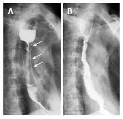
Figure 1 Barium meal examination of the upper gastrointestinal tract.
A: The three metal stents were placed in the esophagus, and the esophageal stricture was on the upper end of the stents. Those three white arrows indicated first, second, and third stent from bottom to top; B: After the removal of all stents, the stricture was alleviated without the occurrence of an esophageal fistula.
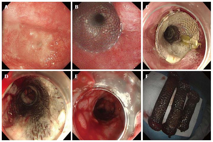
Figure 2 Gastroscope images of esophagus and stents.
Gastroscopy showed (A) an esophageal stricture beginning 25 cm from the incisor teeth; B: The fourth stent was placed and (C) remained in place for 4 wk; D: The previous stents became visible after the removal of the fourth stent; E: Outcomes after the removal of all stents were diffuse, but minor bleeding and mucosal tears, and the imprint of the stent mesh was visible in the esophageal mucosa; F: The three original stents were completely retrieved.
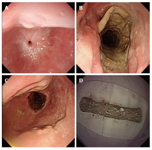
Figure 3 Endoscopic appearances of the esophageal stricture after all the stents were removed.
A: The esophageal stricture located 25 cm from the incisor teeth was worsened 5 wk after the stents were removed, making it difficult to pass the gastroscope; B: A fully covered SEMS was placed and left in place for 4 wk; C: The stricture was significantly wider after the removal of (D) the fully covered SEMS. SEMS: Self-expanding metal stent.
Figure 4 The timeline of stents placement.
- Citation: Liu XQ, Zhou M, Shi WX, Qi YY, Liu H, Li B, Xu HW. Successful endoscopic removal of three embedded esophageal self-expanding metal stents. World J Gastrointest Endosc 2017; 9(9): 494-498
- URL: https://www.wjgnet.com/1948-5190/full/v9/i9/494.htm
- DOI: https://dx.doi.org/10.4253/wjge.v9.i9.494


