Copyright
©The Author(s) 2017.
World J Gastrointest Endosc. Feb 16, 2017; 9(2): 91-94
Published online Feb 16, 2017. doi: 10.4253/wjge.v9.i2.91
Published online Feb 16, 2017. doi: 10.4253/wjge.v9.i2.91
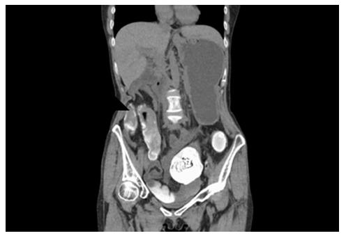
Figure 1 Abdominal computed tomography-scan demonstrating 7 cm fecaloma in the transverse colon.
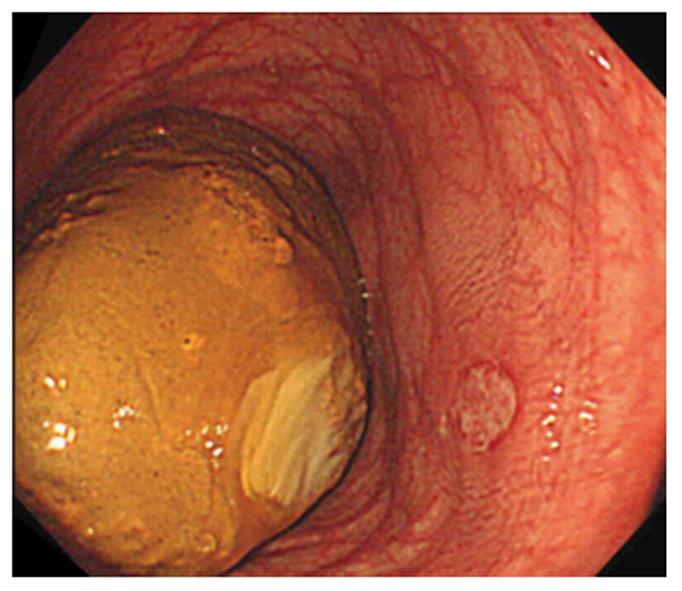
Figure 2 Lower gastrointestinal endoscopy revealed dilated colonic lumen and brown fecaloma in the transverse colon.
There is 2 cm ulcer near the fecaloma.
Figure 3 Same procedure was then repeated several hundred times, aiming for the center of the fecaloma and resulting in gradual fragmentation.
A: Jumbo forceps scrape off the surface of hardened fecaloma; B: Jumbo forceps split the fecaloma; C: Fecaloma is separated into two blocks by biting the jumbo forceps.
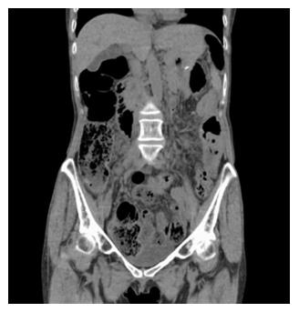
Figure 4 Abdominal computed tomography reveal the disappearance of the fecaloma.
- Citation: Matsuo Y, Yasuda H, Nakano H, Hattori M, Ozawa M, Sato Y, Ikeda Y, Ozawa SI, Yamashita M, Yamamoto H, Itoh F. Successful endoscopic fragmentation of large hardened fecaloma using jumbo forceps. World J Gastrointest Endosc 2017; 9(2): 91-94
- URL: https://www.wjgnet.com/1948-5190/full/v9/i2/91.htm
- DOI: https://dx.doi.org/10.4253/wjge.v9.i2.91


