Copyright
©The Author(s) 2015.
World J Gastrointest Endosc. Jun 25, 2015; 7(7): 747-757
Published online Jun 25, 2015. doi: 10.4253/wjge.v7.i7.747
Published online Jun 25, 2015. doi: 10.4253/wjge.v7.i7.747
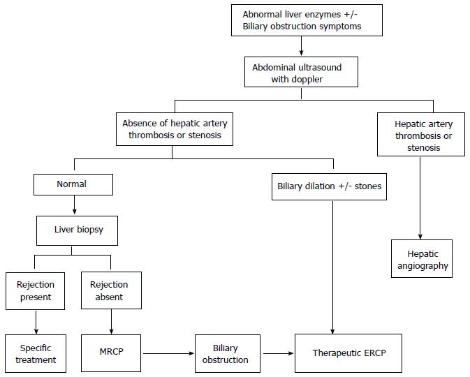
Figure 1 Algorithm for evaluation of suspected biliary obstruction after orthotopic liver transplantation in patients with duct-to-duct reconstruction.
MRCP: Magnetic resonance cholangiopancreatography; ERCP: Endoscopic retrograde cholangiopancreatographys.
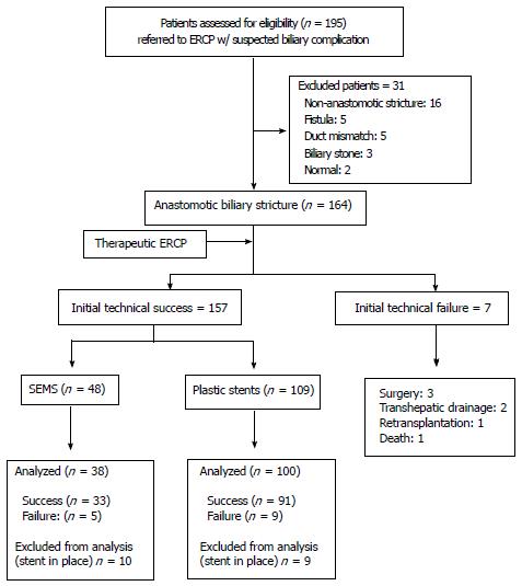
Figure 2 Flow chart of patients in the study.
ERCP: Endoscopic retrograde cholangiopancreatographys; SEMS: Stent-expandable metal stents.
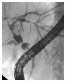
Figure 3 Patient with post-orthotopic liver transplantation anastomotic stricture from index endoscopic retrograde cholangiopancreatographys.
A: Retrograde cholangiogram demonstrating post-OLT anastomotic stricture (arrow); B: Patient was treated with progressive multiple plastic stents; C: Patient was treated with progressive multiple plastic stents; D: Final cholangiogram revealing complete stricture resolution. OLT: Orthotopic liver transplantation.
Figure 4 Patient with post-orthotopic liver transplantation anastomotic stricture.
A: Post-OLT anastomotic biliary stricture; B: Placement of a fully covered SEMS across the stricture as a primary therapy option; C: Endoscopic view of the FCSEMS after 6 mo in place; D: Fluoroscopic image revealing enlargement of the common hepatic duct after SEMS removal. OLT: Orthotopic liver transplantation; FCSEMS: Fully covered stent-expandable metal stents.
Figure 5 Recurrent anastomotic stricture after fully covered stent-expandable metal stents distal migration.
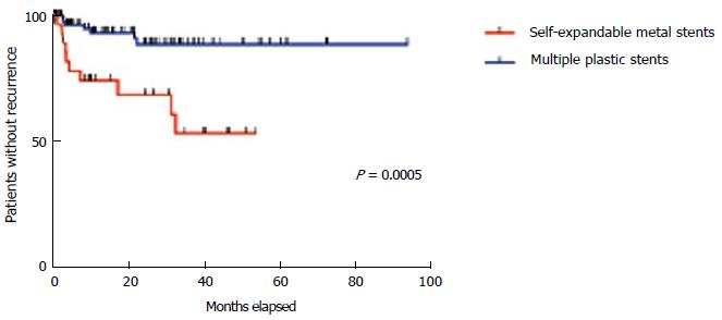
Figure 6 Stricture recurrence after resolution.
- Citation: Martins FP, Kahaleh M, Ferrari AP. Management of liver transplantation biliary stricture: Results from a tertiary hospital. World J Gastrointest Endosc 2015; 7(7): 747-757
- URL: https://www.wjgnet.com/1948-5190/full/v7/i7/747.htm
- DOI: https://dx.doi.org/10.4253/wjge.v7.i7.747


