Copyright
©The Author(s) 2015.
World J Gastrointest Endosc. Oct 25, 2015; 7(15): 1186-1190
Published online Oct 25, 2015. doi: 10.4253/wjge.v7.i15.1186
Published online Oct 25, 2015. doi: 10.4253/wjge.v7.i15.1186
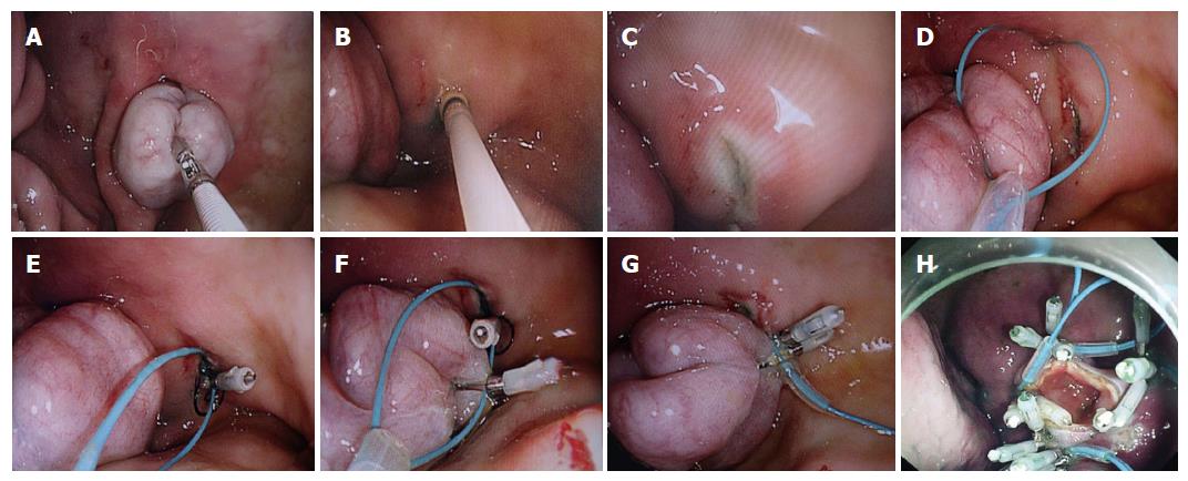
Figure 1 Step-by-step procedure of pure natural orifice transluminal endoscopic surgery side-to-side gastrojejunostomy.
A: Endoscopic view of a loop of the small bowel in the stomach grasped by an endoscopic alligator forceps on its anti-mesenteric side; B: Image taken during submucosal injection around the loop; C: Endoscopic view of one hole made on gastric mucosal surface; D: Endoscopic view of an endoloop placed around the hole; E: Endoscopic view of one clip clipped to anchor the endoloop on the side of the stomach after the prong of the clip was inserted in the hole of the stomach wall; F: Endoscopic view of the second clip clipped to anchor the endoloop on the side of the small intestine; G: Endoscopic view of the endoloop tightened to approximate the gastric mucosal layer and the intestinal serosal layer; H: Endoscopic view of gasto-jejunal mucosal-seromuscular layer anastomosis followed by the loop full-thickness incision.
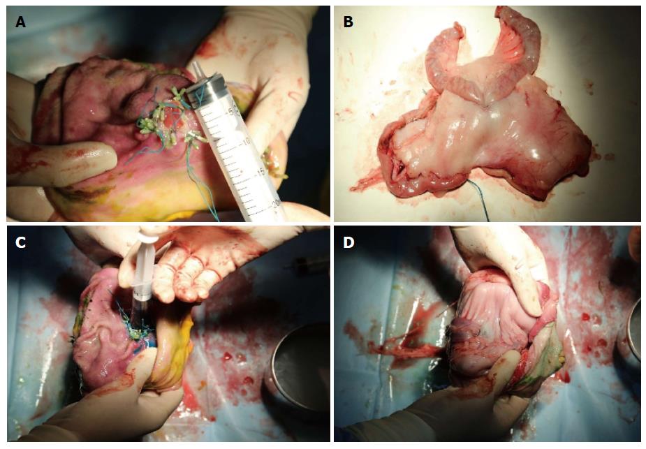
Figure 2 Postmortem appearances of anastomosis.
A: Macroscopic appearance showing that the intestinal wall had been joined to the stomach wall; B: Macroscopic appearance of gastrointestinal side-to-side anastomosis; C: Methylene blue instilled into the anastomotic lumen; D: No methylene blue observed on the surface of gastric serosa around the anastomosis.
- Citation: Chen SY, Shi H, Jiang SJ, Wang YG, Lin K, Xie ZF, Liu XJ. Transgastric endoscopic gastrojejunostomy using holing followed by interrupted suture technique in a porcine model. World J Gastrointest Endosc 2015; 7(15): 1186-1190
- URL: https://www.wjgnet.com/1948-5190/full/v7/i15/1186.htm
- DOI: https://dx.doi.org/10.4253/wjge.v7.i15.1186


