Copyright
©The Author(s) 2015.
World J Gastrointest Endosc. Sep 10, 2015; 7(12): 1070-1077
Published online Sep 10, 2015. doi: 10.4253/wjge.v7.i12.1070
Published online Sep 10, 2015. doi: 10.4253/wjge.v7.i12.1070
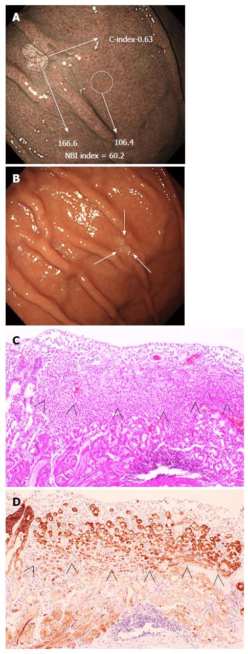
Figure 1 Endoscopic and histologic findings of signet ring cell carcinomas localized at the greater curvature of the corpus (Case 4).
A: NBI of the cancer area showed an isolated oval-shaped whitish area. The NBI index was 60.2. The C-index was 0.63; B: Endoscopy with WLI showed a 0-IIc lesion with slight discoloration (arrows) at the greater curvature of the corpus. The WLI index was 4.5; C: The histology by endoscopic submucosal dissection showed an intramucosal SRC in the upper third of the gastric mucosa (arrowheads) with a partial defect of the foveolar epithelium; D: Immunohistochemical staining of SRC cells showed diffuse positive reactivity for cytokeratin AE1/AE3 (arrowheads). NBI: Narrow-band imaging; WLI: White-light imaging; SRC: Signet ring cell carcinoma.
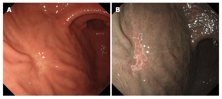
Figure 2 Endoscopic images of whitish gastric ulcer scars.
White-light imaging (A) and non-magnifying narrow-band imaging (B). The NBI index and C-index were 44.9 and 0.29, respectively. NBI: Narrow-band imaging; C-index: Circularity index.
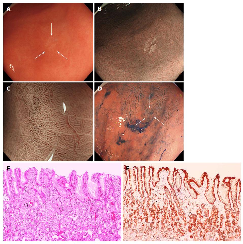
Figure 3 Endoscopic and microscopic images of signet ring cell carcinoma (Case 9).
A: Endoscopy with WLI revealed a 0-IIb lesion with slight discoloration (arrows) at the greater curvature of the angulus; B: NM-NBI of the cancer area showed an isolated clear whitish area. The cancerous areas were more clearly captured by NM-NBI endoscopy than by WLI endoscopy. The NBI and WLI index were respectively 25.6 and 12.8, and the C-index was 0.77; C and D: The demarcation line of the SRC was not clearly identified even by magnifying NBI and chromoendoscopy (arrows); E: The histology of a specimen resected by endoscopic submucosal dissection revealed an intramucosal SRC (arrowheads) in the upper third of the mucosa beneath a preserved surface epithelium; F: SRC cells showed positive for cytokeratin AE1/AE3 staining (arrowheads). WLI: White-light imaging; NM-NBI: Non-magnifying narrow-band imaging; SRC: Signet ring cell carcinoma.
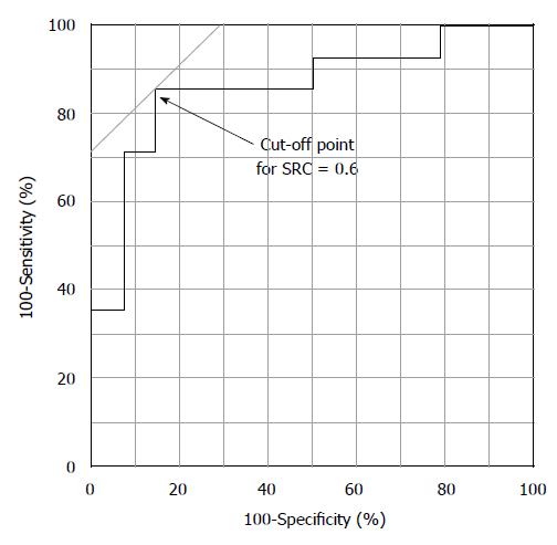
Figure 4 Receiver operating characteristic curve of the C-index of signet ring cell carcinoma.
The curve is plotted as sensitivity (Y axis) and (100- specificity) (X axis). SRC: Signet ring cell carcinoma.
- Citation: Watari J, Tomita T, Ikehara H, Taki M, Ogawa T, Yamasaki T, Kondo T, Toyoshima F, Sakurai J, Kono T, Tozawa K, Ohda Y, Oshima T, Fukui H, Hirota S, Miwa H. Diagnosis of small intramucosal signet ring cell carcinoma of the stomach by non-magnifying narrow-band imaging: A pilot study. World J Gastrointest Endosc 2015; 7(12): 1070-1077
- URL: https://www.wjgnet.com/1948-5190/full/v7/i12/1070.htm
- DOI: https://dx.doi.org/10.4253/wjge.v7.i12.1070


