Copyright
©The Author(s) 2015.
World J Gastrointest Endosc. Jan 16, 2015; 7(1): 73-76
Published online Jan 16, 2015. doi: 10.4253/wjge.v7.i1.73
Published online Jan 16, 2015. doi: 10.4253/wjge.v7.i1.73
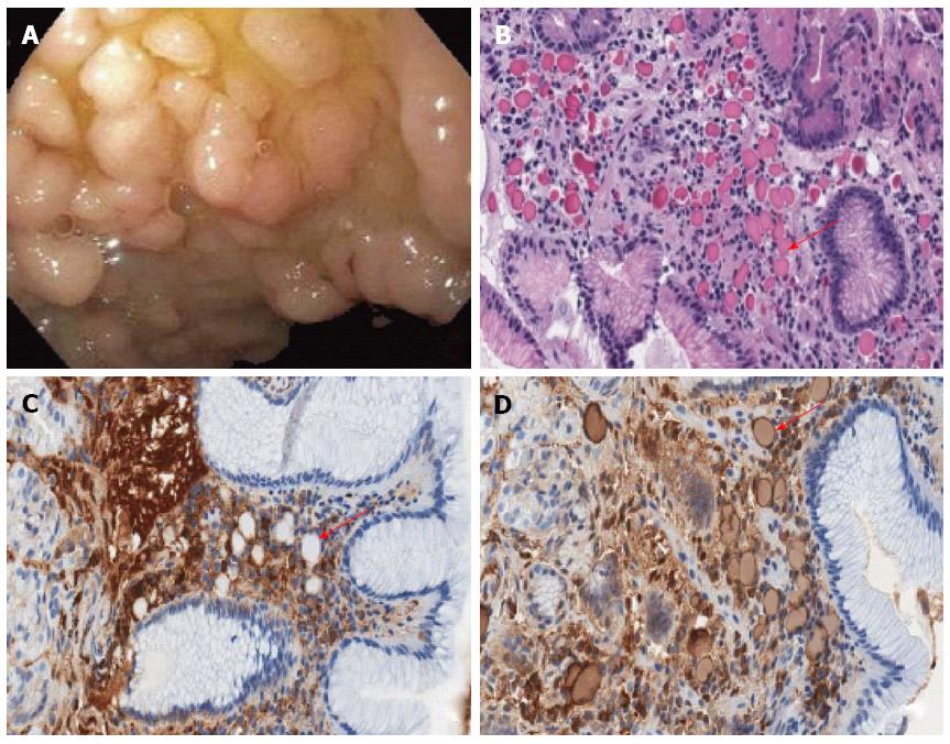
Figure 1 Endoscopic findings of duodenum.
A: Endoscopic image shows a cluster of lobulated polyps in duodenal bulb; B: Numerous eosinophilic inclusions (Russell bodies, arrow) in the lamina propria and gastric foveolar metaplasia of the duodenal surface epithelium. H and E, 400 ×; C: CD138 immunohistochemistry highlights numerous mature plasma cells. The Russell bodies are round to ovoid clear spaces (arrow) within the lamina propria. Immunohistochemistry, 400 ×; D: Immunoglobulin kappa light chain immunohistochemistry shows a dark-rim staining and light internal staining pattern of Russell bodies (arrow). Immunohistochemistry, 400 ×.
- Citation: Munday WR, Kapur LH, Xu M, Zhang X. Russell body duodenitis with immunoglobulin kappa light chain restriction. World J Gastrointest Endosc 2015; 7(1): 73-76
- URL: https://www.wjgnet.com/1948-5190/full/v7/i1/73.htm
- DOI: https://dx.doi.org/10.4253/wjge.v7.i1.73


