Copyright
©2013 Baishideng Publishing Group Co.
World J Gastrointest Endosc. Sep 16, 2013; 5(9): 461-464
Published online Sep 16, 2013. doi: 10.4253/wjge.v5.i9.461
Published online Sep 16, 2013. doi: 10.4253/wjge.v5.i9.461
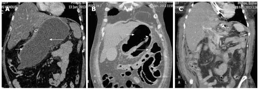
Figure 1 Computerized Tomography of abdomen showing pseudocyst of pancreas.
A: Large pseudocyst (long arrow) causing extrinsic compression of stomach (short arrow); B: Ruptured pseudocyst of pancreas (long arrow) draining through a rent (arrowhead) into stomach (short arrow); C: Complete resolution of pseudocyst of pancreas.
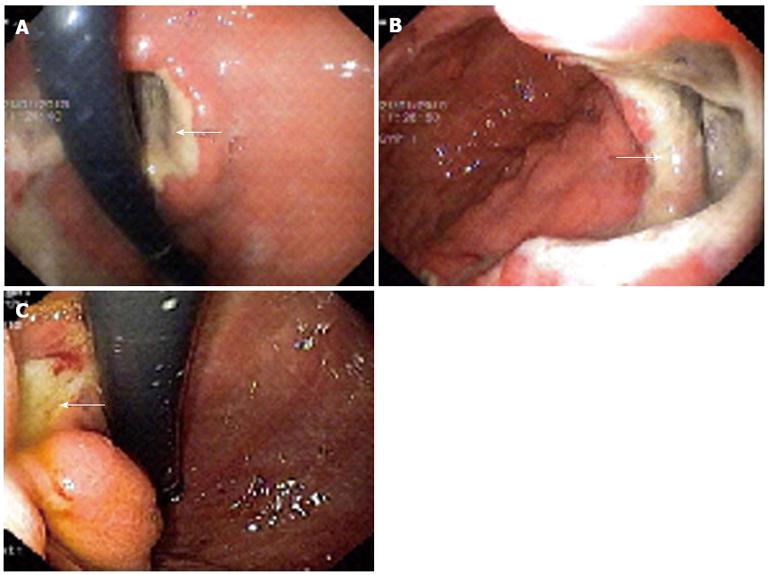
Figure 2 Oesophagogastroduodenoscopy showing pseudocyst of pancreas.
A, B: Orifice (arrow) of the fistula between stomach and the pseudocyst of pancreas; C: Ulcer (arrow) at the site of healed fistulous communication between pancreatic pseudocyst and stomach.
- Citation: Somani PO, Jain SS, Shah DK, Khot AA, Rathi PM. Uncomplicated spontaneous rupture of pancreatic pseudocyst into stomach: A case report. World J Gastrointest Endosc 2013; 5(9): 461-464
- URL: https://www.wjgnet.com/1948-5190/full/v5/i9/461.htm
- DOI: https://dx.doi.org/10.4253/wjge.v5.i9.461


