Copyright
©2013 Baishideng Publishing Group Co.
World J Gastrointest Endosc. Sep 16, 2013; 5(9): 457-460
Published online Sep 16, 2013. doi: 10.4253/wjge.v5.i9.457
Published online Sep 16, 2013. doi: 10.4253/wjge.v5.i9.457
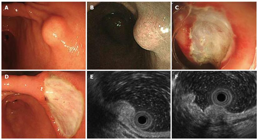
Figure 1 Endoscopic ultrasonography findings of visualized submucosal tumor.
A: Endoscopy shows 10-mm shows submucosal tumor (SMT) in lesser curvature of lower corpus of stomach; B: Narrow-band endoscopic imaging SMT covered by normal gastric mucosa; C: Endoscopic submucosal dissection (ESD) for SMT; D: Stomach ulceration five days after ESD; E: Endoscopic ultrasonography (EUS) findings show internally isoechoic, homogeneous sub-mucosal tumor mainly localized within second and third layers, whereas first and fourth layers are preserved; F: Acoustic shadowing of hyperechoic foci inside lesion is consistent with calcifications. Fourth layer is obvious.
Figure 2 Pathological findings of resected small mucosal tumor.
A: Complete submucosal tumor resection was confirmed; B: Hypocellular, spindle-cell tumor has densely hyalinized, collagenous matrix, scattered lymphoplasmacytic aggregates, some psammomatous foci and dystrophic calcification, Spindle-cells harbor no mitotic activity or atypia; C: Positive immunohistochemical staining for vimentin. Original magnification × 10 (A), × 200 (B), × 100 (C).
- Citation: Ogasawara N, Izawa S, Mizuno M, Tanabe A, Ozeki T, Noda H, Takahashi E, Sasaki M, Yokoi T, Kasugai K. Gastric calcifying fibrous tumor removed by endoscopic submucosal dissection. World J Gastrointest Endosc 2013; 5(9): 457-460
- URL: https://www.wjgnet.com/1948-5190/full/v5/i9/457.htm
- DOI: https://dx.doi.org/10.4253/wjge.v5.i9.457


