Copyright
©2013 Baishideng Publishing Group Co.
World J Gastrointest Endosc. Mar 16, 2013; 5(3): 138-140
Published online Mar 16, 2013. doi: 10.4253/wjge.v5.i3.138
Published online Mar 16, 2013. doi: 10.4253/wjge.v5.i3.138
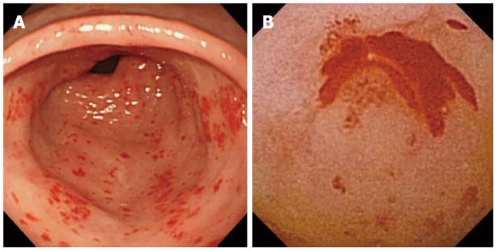
Figure 1 Conventional and capsule endoscopy.
A: Conventional endoscopy showing non-bleeding gastric antral vascular ectasia (GAVE), mislabeled as superficial gastritis; B: Capsule endoscopy showing active bleeding from GAVE.
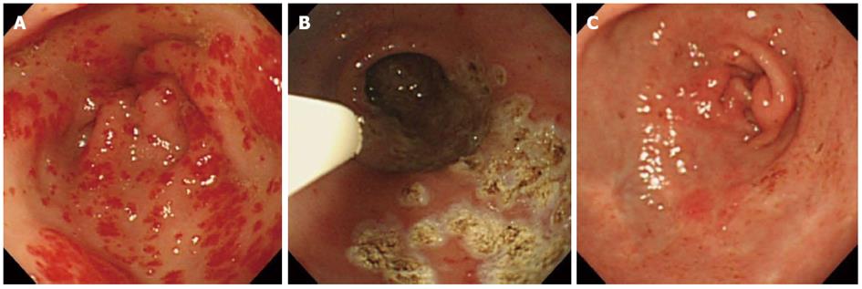
Figure 2 Endoscopy showing gastric antral vascular ectasia.
A: Endoscopy showing gastric antral vascular ectasia (GAVE) with the classical “watermelon stomach” appearance just before the treatment with argon plasma coagulation (APC); B: Endoscopy showing GAVE during the treatment with APC; C: Endoscopy showing the improvement of GAVE with scar formation.
- Citation: Ohira T, Hokama A, Kinjo N, Nakamoto M, Kobashigawa C, Kise Y, Yamashiro S, Kinjo F, Kuniyoshi Y, Fujita J. Detection of active bleeding from gastric antral vascular ectasia by capsule endoscopy. World J Gastrointest Endosc 2013; 5(3): 138-140
- URL: https://www.wjgnet.com/1948-5190/full/v5/i3/138.htm
- DOI: https://dx.doi.org/10.4253/wjge.v5.i3.138


