Copyright
©2010 Baishideng.
World J Gastrointest Endosc. Feb 16, 2010; 2(2): 61-68
Published online Feb 16, 2010. doi: 10.4253/wjge.v2.i2.61
Published online Feb 16, 2010. doi: 10.4253/wjge.v2.i2.61
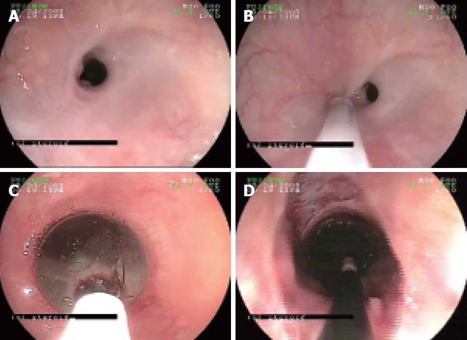
Figure 1 Endoscopic image of an esophageal stricture and its management.
A: A refractory stricture; B: Steroid injection at the proximal edge of the stricture; C: Through-the-scope (TTS) balloon dilation of the stricture; D: The opened stricture after deflation of the TTS balloon.
Figure 2 Barium swallow image of a stricture.
A: A complex resistant esophageal stricture; B: The opened stricture after intralesional steroid injection and balloon dilation.
- Citation: Kochhar R, Poornachandra KS. Intralesional steroid injection therapy in the management of resistant gastrointestinal strictures. World J Gastrointest Endosc 2010; 2(2): 61-68
- URL: https://www.wjgnet.com/1948-5190/full/v2/i2/61.htm
- DOI: https://dx.doi.org/10.4253/wjge.v2.i2.61


