Copyright
©The Author(s) 2024.
World J Gastrointest Endosc. Jun 16, 2024; 16(6): 361-367
Published online Jun 16, 2024. doi: 10.4253/wjge.v16.i6.361
Published online Jun 16, 2024. doi: 10.4253/wjge.v16.i6.361
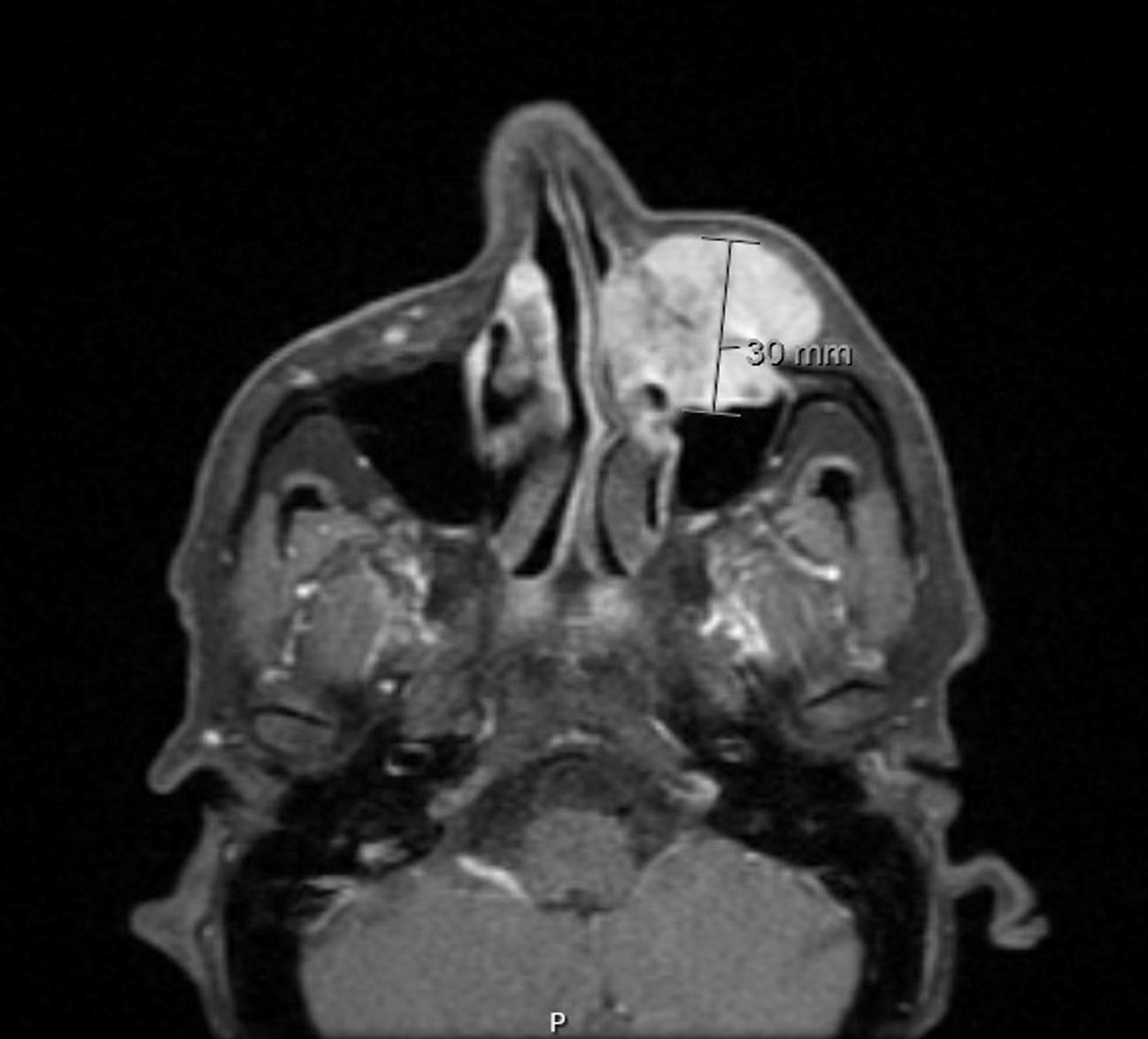
Figure 1 Magnetic resonance imaging of the face and neck showing a left maxillary mass.
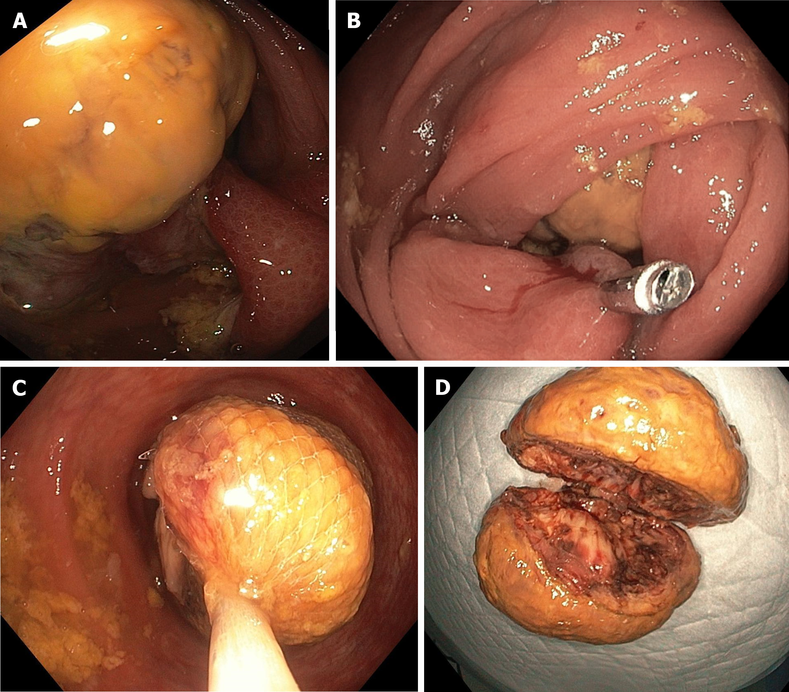
Figure 2 Colonoscopy.
A: Ascending colon polyp; B: Hemoclip applied to ascending colon polyp; C: Roth net covering ascending colon polyp; D: Retrieved ascending colon polyp.
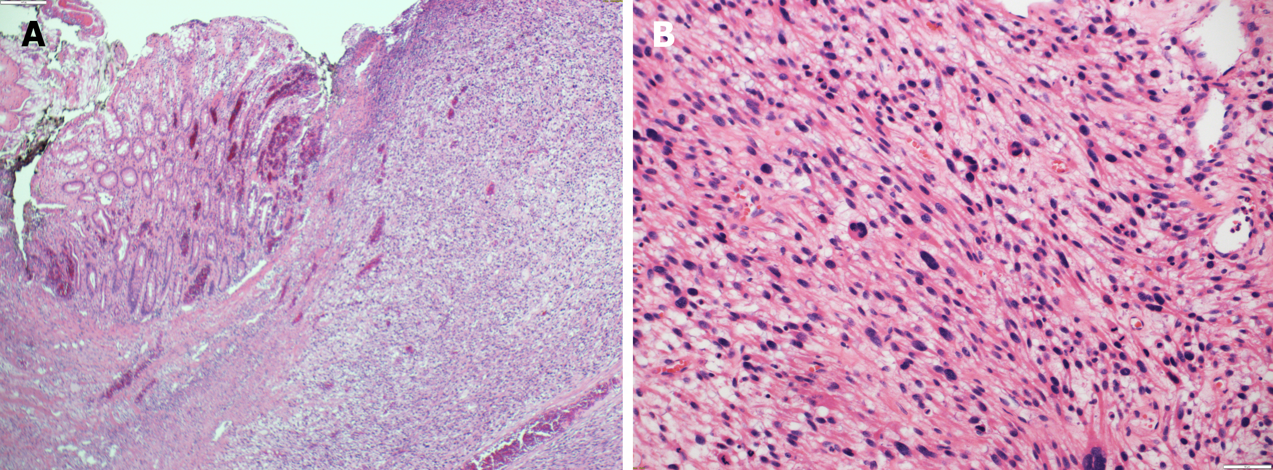
Figure 3 Histopathological examination.
A: Submucosal diffuse spindle cell neoplasm [hematoxylin and eosin (H&E) × 10]; B: Higher magnification of the spindle cell neoplasm showed high mitotic figures and prominent nuclear pleomorphism (H&E × 20).
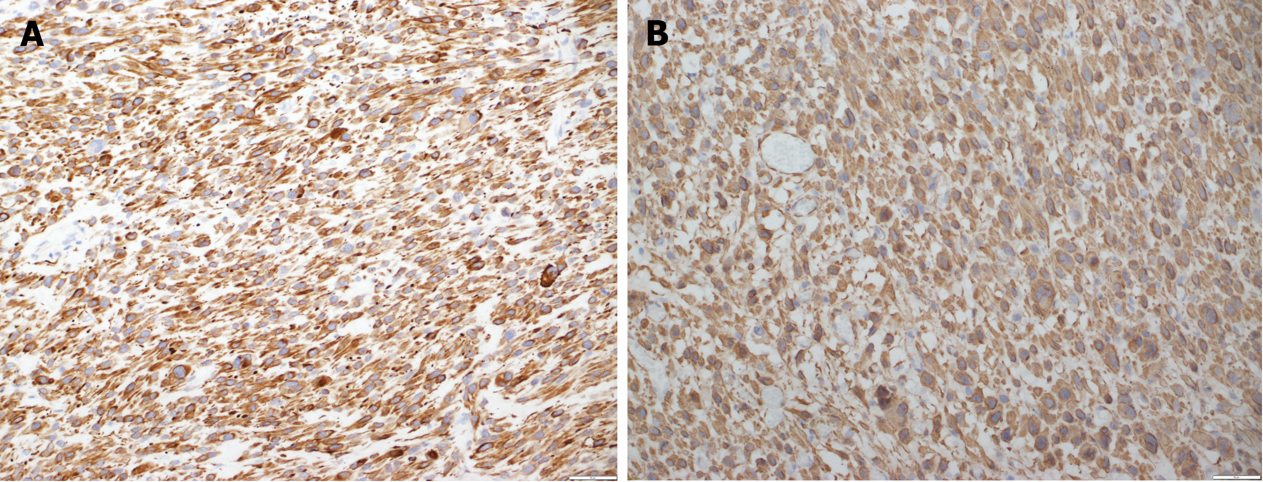
Figure 4 Immunohistochemistry.
A: The malignant cells are positive with desmin [immunohistochemistry (IHC) × 20]; B: The malignant cells are positive with SMA (IHC × 20).
- Citation: Alnajjar A, Alfadda A, Alqaraawi AM, Alajlan B, Atallah JP, AlHussaini HF. Pleomorphic leiomyosarcoma of the maxilla with metastasis to the colon: A case report. World J Gastrointest Endosc 2024; 16(6): 361-367
- URL: https://www.wjgnet.com/1948-5190/full/v16/i6/361.htm
- DOI: https://dx.doi.org/10.4253/wjge.v16.i6.361


