Copyright
©The Author(s) 2023.
World J Gastrointest Endosc. Apr 16, 2024; 16(4): 206-213
Published online Apr 16, 2024. doi: 10.4253/wjge.v16.i4.206
Published online Apr 16, 2024. doi: 10.4253/wjge.v16.i4.206
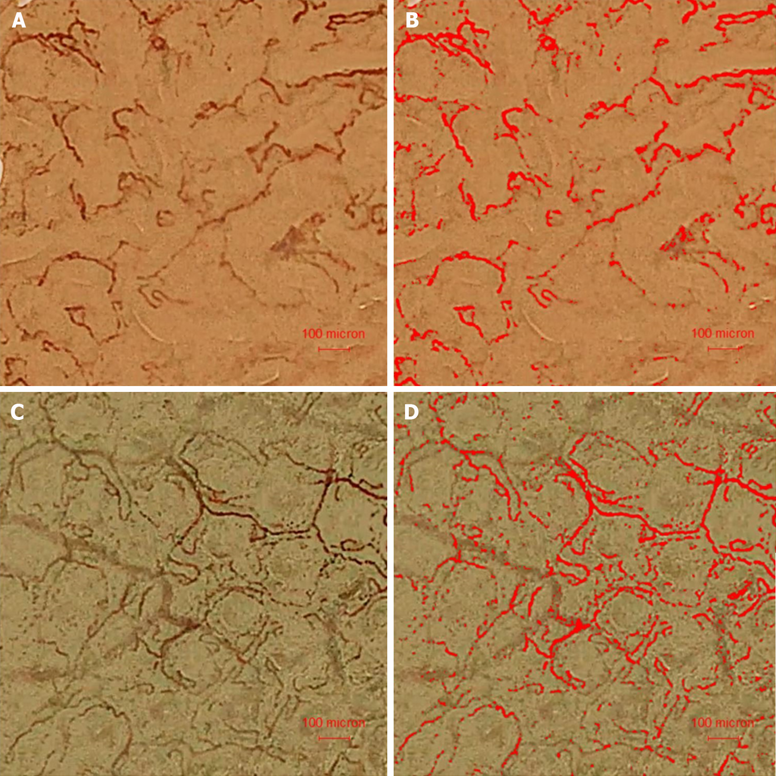
Figure 1 Superficial capillaries in an adenoma and surrounding normal mucosa observed under high-resolution magnifying endoscope with blue-laser imaging, and then identified by Image-pro plus software.
A: Blue laser imaging (BLI) observed the surface vessels of adenomas; B: BLI observed normal mucosal surface vessels around the adenoma; C: Image-pro plus identified the blood vessels on the surface of adenoma as red; D: Image-pro plus identified normal mucosal surface blood vessels around the adenoma as red. BLI: Blue-laser imaging.
Figure 2 The green marks represent the blood flow progression in 1/30 s.
A: The green mark represents the blood flow position at a certain time; B: The green mark represents the blood flow position after 1/30 s.
- Citation: Dong HB, Chen T, Zhang XF, Ren YT, Jiang B. In vivo pilot study into superficial microcirculatory characteristics of colorectal adenomas using novel high-resolution magnifying endoscopy with blue laser imaging. World J Gastrointest Endosc 2024; 16(4): 206-213
- URL: https://www.wjgnet.com/1948-5190/full/v16/i4/206.htm
- DOI: https://dx.doi.org/10.4253/wjge.v16.i4.206


