Copyright
©The Author(s) 2023.
World J Gastrointest Endosc. Feb 16, 2023; 15(2): 19-31
Published online Feb 16, 2023. doi: 10.4253/wjge.v15.i2.19
Published online Feb 16, 2023. doi: 10.4253/wjge.v15.i2.19
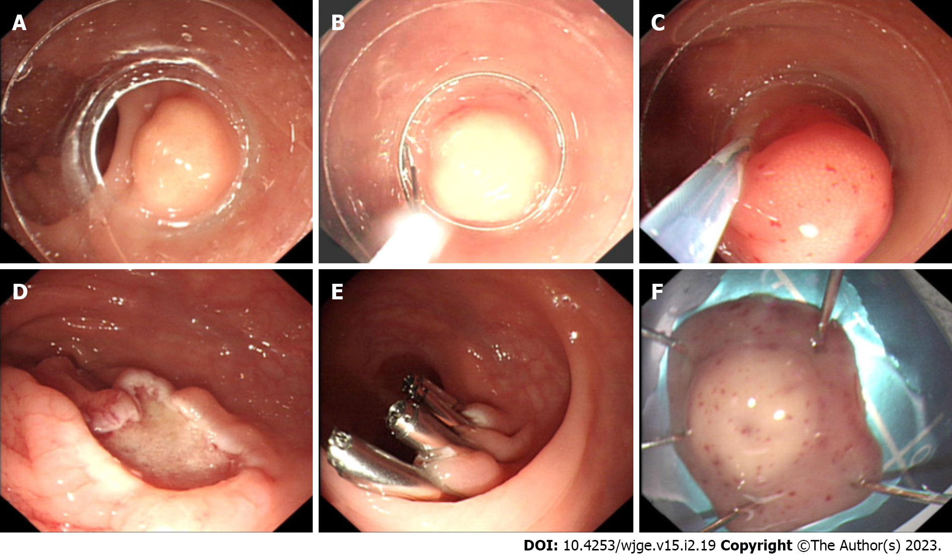
Figure 1 Cap-assisted endoscopic mucosal resection.
A: A pale yellow mass with a diameter of about 9 mm in the rectum; B: Placement of an endoscope with a transparent cap worn at its front end into a crescent snare; C: Resection of the mass with the crescent snare after negative pressure suction; D: Wound surface after the removal of the mass; E: Wound clipping with titanium clips; F: The resected mass for pathological biopsy.
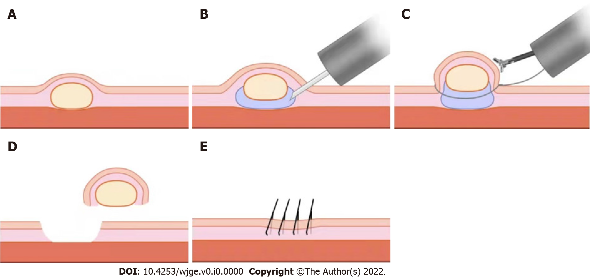
Figure 2 Endoscopic mucosal resection using a dual-channel endoscope.
A: A pale yellow mass in the rectum; B: Submucosal injection of the mass with an injection needle; C: The use of a dual-channel endoscope, with one channel inserted with forceps to lift the lesion, and the other inserted with an electrosurgical snare to resect the mass; D and E: Wound clipping with titanium clips after mass resection.
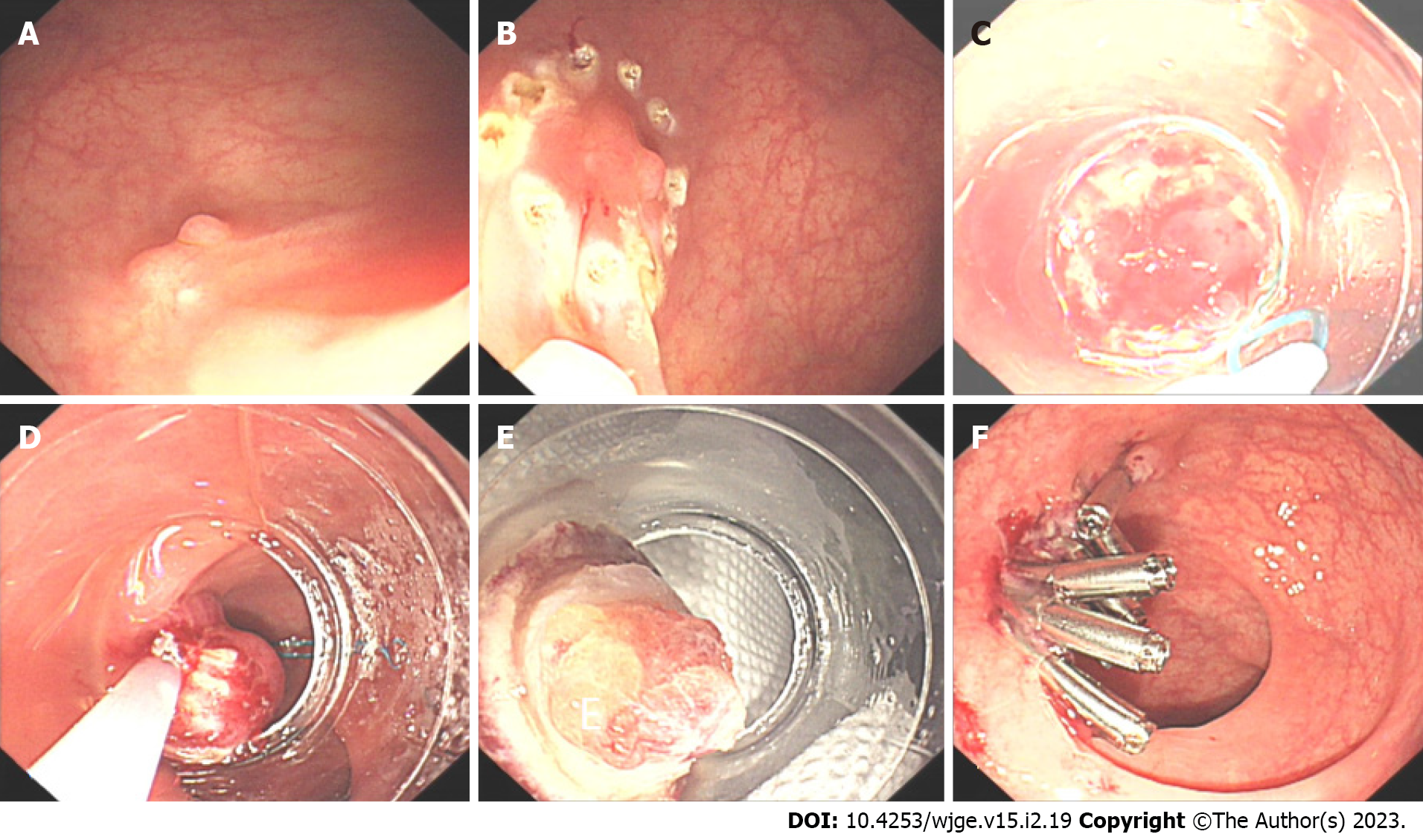
Figure 3 Endoscopic mucosal resection with a ligation device.
A: A pale yellow mass with a diameter of about 6 mm in the rectum, with visible scar after biopsy on the surface; B: Electrocoagulation marking in the peritumoral area by using the front end of the electrosurgical snare; C: Ligation of the root of the mass after negative pressure suction with a single-ring nylon ring; D: Resection of the mass at the root with an electrosurgical snare; E: Resected mass; F: Wound clipping with titanium clips after mass resection.
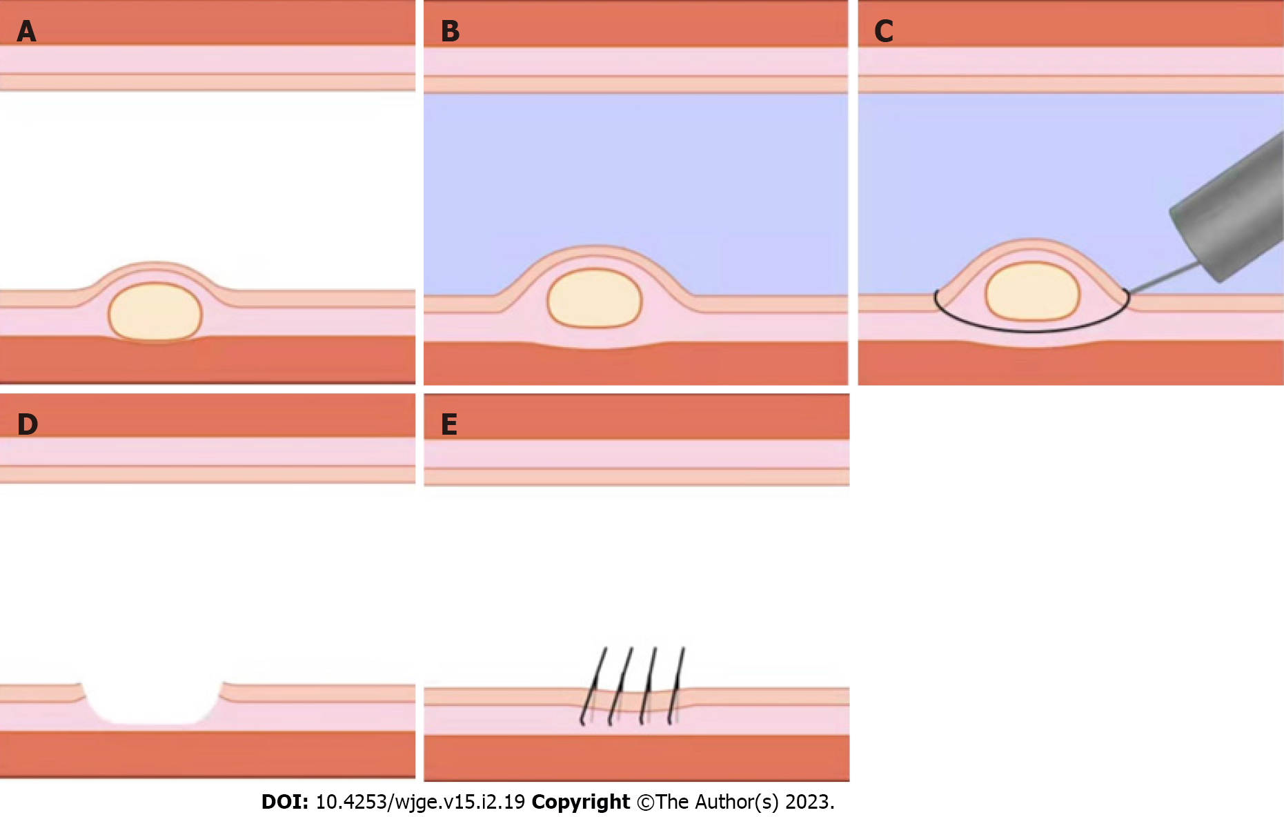
Figure 4 Underwater endoscopic mucosal resection.
A: A pale yellow mass in the rectum; B: Floating of the mass through the buoyancy of water after air extraction and water injection into the rectum; C: Resection of the mass by using electrosurgical snare; D and E: Wound clipping with titanium clips after mass resection.
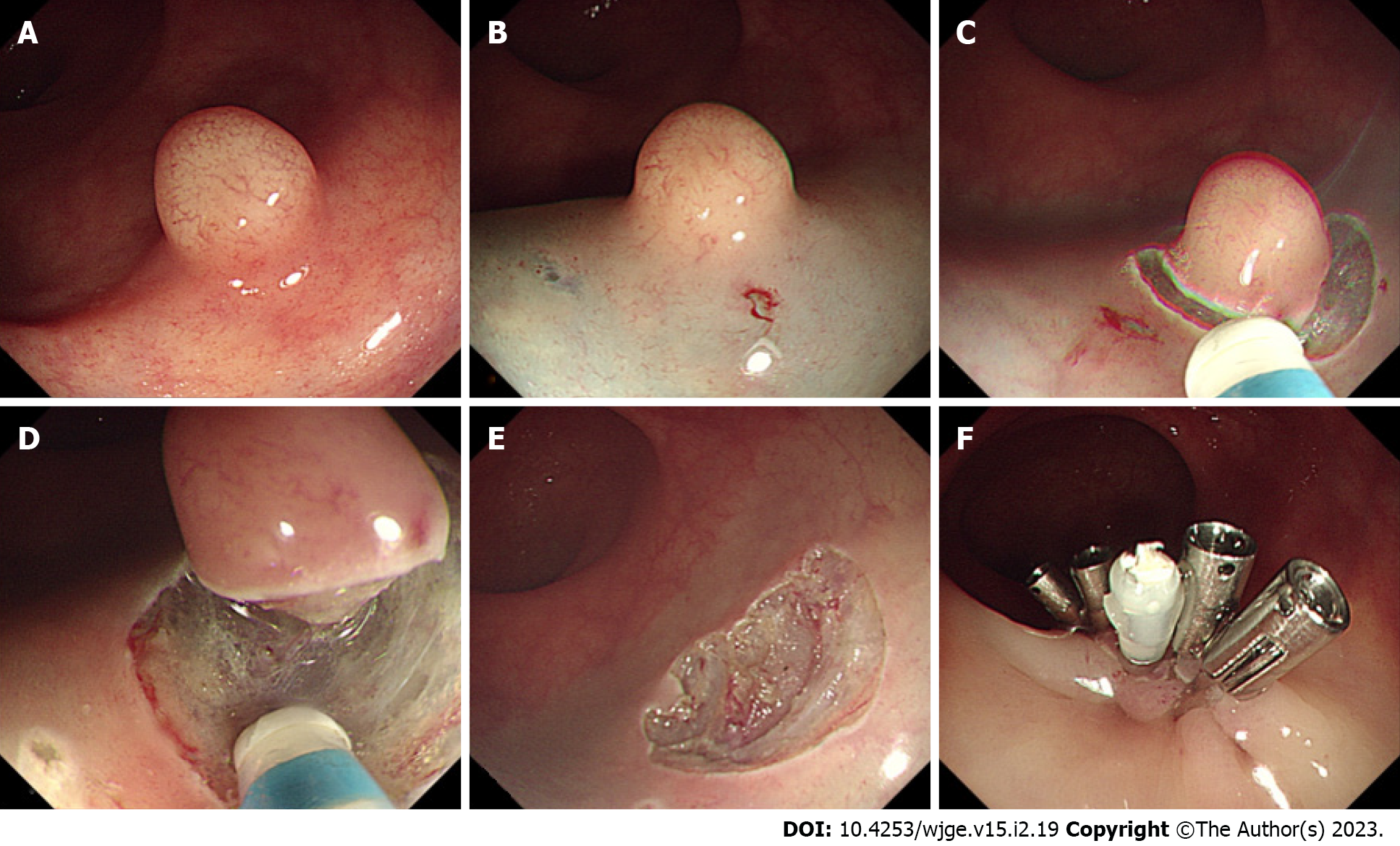
Figure 5 Endoscopic submucosal dissection.
A: A pale yellow mass with a diameter of about 6 mm in the rectum; B: Submucosal injection of the mass with an injection needle; C: Circumferential resection of the submucosa of the mass with a mucosal resection knife; D: Mass dissection with a resection knife; E: Wound surface after the removal of the mass; F: Wound clipping with titanium clips after mass resection.
- Citation: Ma XX, Wang LS, Wang LL, Long T, Xu ZL. Endoscopic treatment and management of rectal neuroendocrine tumors less than 10 mm in diameter. World J Gastrointest Endosc 2023; 15(2): 19-31
- URL: https://www.wjgnet.com/1948-5190/full/v15/i2/19.htm
- DOI: https://dx.doi.org/10.4253/wjge.v15.i2.19


