Copyright
©The Author(s) 2022.
World J Gastrointest Endosc. Mar 16, 2022; 14(3): 129-141
Published online Mar 16, 2022. doi: 10.4253/wjge.v14.i3.129
Published online Mar 16, 2022. doi: 10.4253/wjge.v14.i3.129
Figure 1 Case No.
13: A 77 years old woman with a pancreatic cyst lesion corresponding to an intraductal papillary mucinous neoplasm. The lesion exhibited malignancy criteria at endoscopic ultrasound (EUS) and related techniques. A: EUS identifying a 4 cm pancreatic cyst lesion with mural nodules (yellow arrow); B: Mural nodule with hyper-enhancing at EUS (green arrow) shown in contrast-enhanced EUS; C: EUS-guided cystoscopy using a digital probe showing vascularity (red arrow) of a pancreatic macrocystic lesion filled with clear fluid.
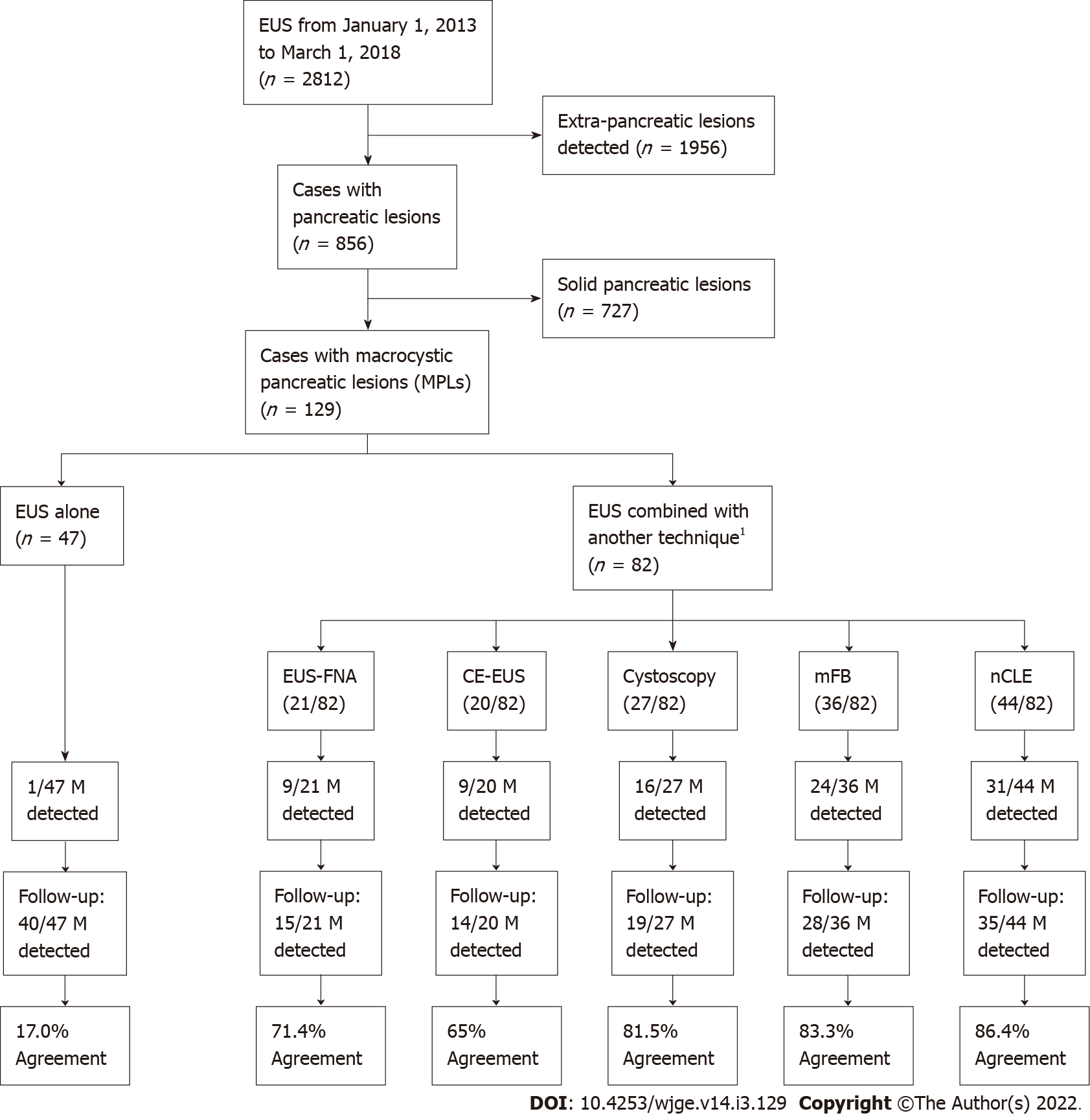
Figure 2 Population study flowchart.
1Numbers of techniques were not mutually exclusive. Endoscopic ultrasound could be combined with more than one other technique, as shown on the illustrated Venn diagram in Figure 3. EUS: Endoscopic ultrasound; EUS-FNA: Endoscopic ultrasound-guided fine needle aspiration; Cystoscopy: Fiberoptic probe cystoscopy; nCLE: Endoscopic ultrasound-guided needle-based confocal laser-endomicroscopy; mFB: Endoscopic ultrasound-guided through-the-needle direct intracystic micro forceps biopsy; CE-EUS: Contrast-enhanced endoscopic ultrasound; M: Malignancy.
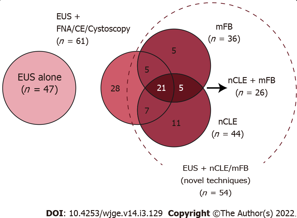
Figure 3 Venn diagram describing distribution of additional diagnostic techniques performed in the studied population.
EUS: Endoscopic ultrasound; EUS-FNA: Endoscopic ultrasound-guided fine needle aspiration; Cystoscopy: Fiberoptic probe cystoscopy; nCLE: Endoscopic ultrasound-guided needle-based confocal laser-endomicroscopy; mFB: Endoscopic ultrasound-guided through-the-needle direct intracystic micro forceps biopsy; CE-EUS: Contrast-enhanced endoscopic ultrasound.
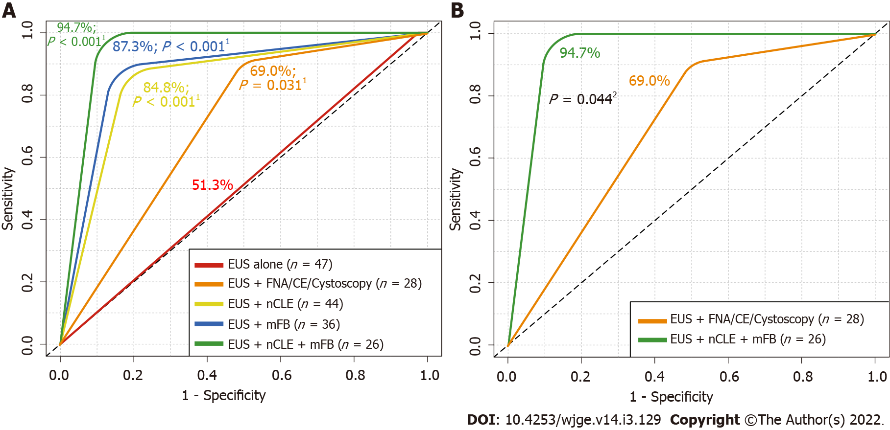
Figure 4 Received operating characteristics describing overall diagnostic accuracy of endoscopic ultrasound alone and in addition with fine needle aspiration or contrast-enhanced endoscopic ultrasound, needle-based confocal laser-endomicroscopy and/or with direct intracystic micro forceps biopsy for detecting malignancy.
A: Comparison among endoscopic ultrasound (EUS) alone vs additional diagnostic techniques; B: Comparison among EUS alone vs EUS + EUS-guided needle-based confocal laser-endomicroscopy (nCLE) + EUS-guided through-the-needle direct intracystic micro forceps biopsy (mFB). 1DeLong’s test for two received operating characteristics (ROC) curves comparing EUS-alone area under the ROC curve (red line) with EUS + fine needle aspiration (FNA)/contrast-enhanced (CE) (orange line), EUS + nCLE (yellow line), EUS + mFB (blue line) and EUS + nCLE + mFB (green line). 2DeLong’s test for two ROC curves comparing EUS + FNA/CE (orange line) with EUS + nCLE + mFB (green line). EUS: Endoscopic ultrasound; FNA: Fine needle aspiration; Cystoscopy: Fiberoptic probe cystoscopy; nCLE: Endoscopic ultrasound-guided needle-based confocal laser-endomicroscopy; mFB: Endoscopic ultrasound-guided through-the-needle direct intracystic micro forceps biopsy; CE: Contrast-enhanced.
- Citation: Robles-Medranda C, Olmos JI, Puga-Tejada M, Oleas R, Baquerizo-Burgos J, Arevalo-Mora M, Del Valle Zavala R, Nebel JA, Calle Loffredo D, Pitanga-Lukashok H. Endoscopic ultrasound-guided through-the-needle microforceps biopsy and needle-based confocal laser-endomicroscopy increase detection of potentially malignant pancreatic cystic lesions: A single-center study. World J Gastrointest Endosc 2022; 14(3): 129-141
- URL: https://www.wjgnet.com/1948-5190/full/v14/i3/129.htm
- DOI: https://dx.doi.org/10.4253/wjge.v14.i3.129


