Copyright
©The Author(s) 2022.
World J Gastrointest Endosc. Oct 16, 2022; 14(10): 636-641
Published online Oct 16, 2022. doi: 10.4253/wjge.v14.i10.636
Published online Oct 16, 2022. doi: 10.4253/wjge.v14.i10.636
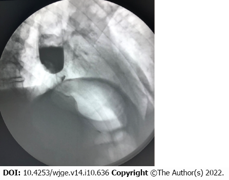
Figure 1 X-ray of esophagus revealing a filling defect.
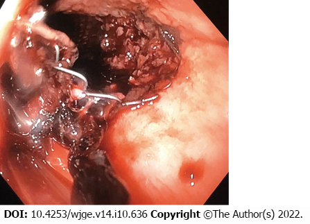
Figure 2 Endoscopic view of bleeding with visualization of uncovered part of the stent.
There were visible signs of tumor growth.
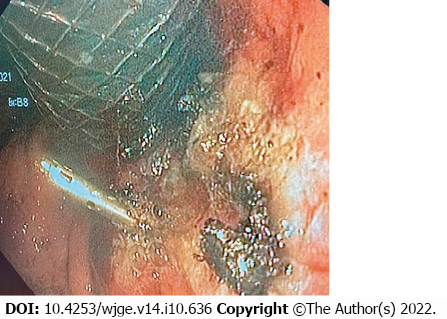
Figure 3 View of a clot in the gastroesophageal junction after stent removal.
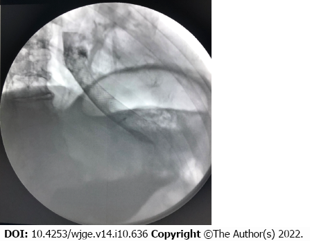
Figure 4 X-ray of esophagus.
Correct location of the stent in the gastroesophageal junction was visualized.
- Citation: Kashintsev AA, Rusanov DS, Antipova MV, Anisimov SV, Granstrem OK, Kokhanenko NY, Medvedev KV, Kutumov EB, Nadeeva AA, Proutski V. Hemostasis of massive bleeding from esophageal tumor: A case report. World J Gastrointest Endosc 2022; 14(10): 636-641
- URL: https://www.wjgnet.com/1948-5190/full/v14/i10/636.htm
- DOI: https://dx.doi.org/10.4253/wjge.v14.i10.636


