Copyright
©The Author(s) 2021.
World J Gastrointest Endosc. Oct 16, 2021; 13(10): 491-501
Published online Oct 16, 2021. doi: 10.4253/wjge.v13.i10.491
Published online Oct 16, 2021. doi: 10.4253/wjge.v13.i10.491
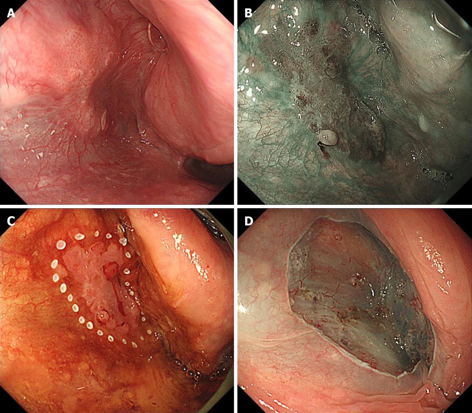
Figure 1 A case of T1 hypopharyngeal cancer located in the left pyriform sinus, detected by gastrointestinal endoscopy.
A: The lesion was recognized as a slightly reddish area under white light image endoscopy; B: The lesion was clearly visualized using narrow-band imaging; C, D: Under general anesthesia, en bloc endoscopic submucosal dissection was successfully completed.
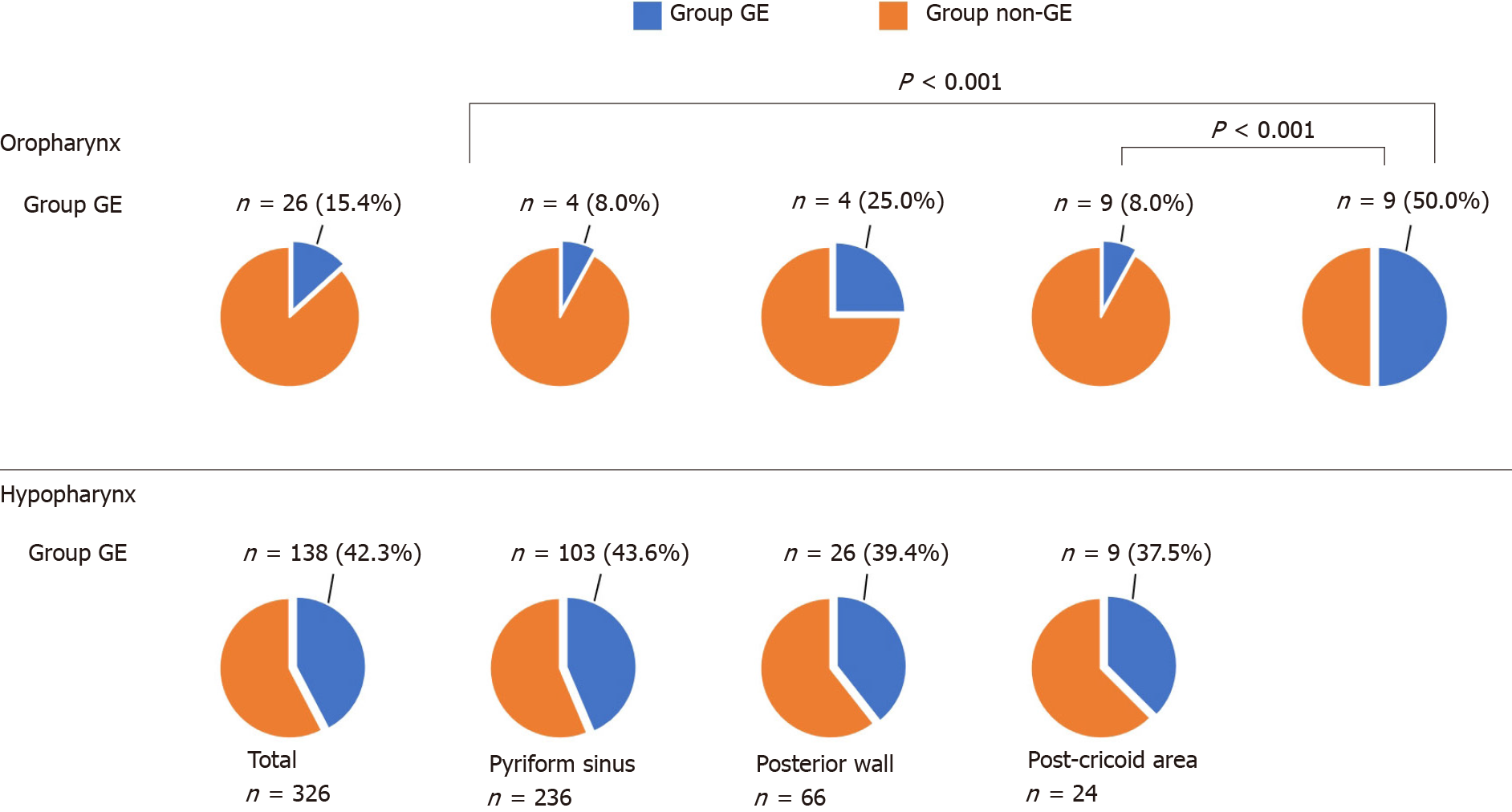
Figure 2 The subsites of primary lesions and the proportion of Group gastrointestinal endoscopy by subsite.
The proportions of Group gastrointestinal endoscopy (GE) in the oropharynx and hypopharynx were 15.4% and 42.3%, respectively. Among the lesions in the oropharynx, the proportions of Group GE in the anterior and lateral wall were lower than the posterior wall. There was no significant difference in the proportion of Group GE by subsite in the hypopharynx.
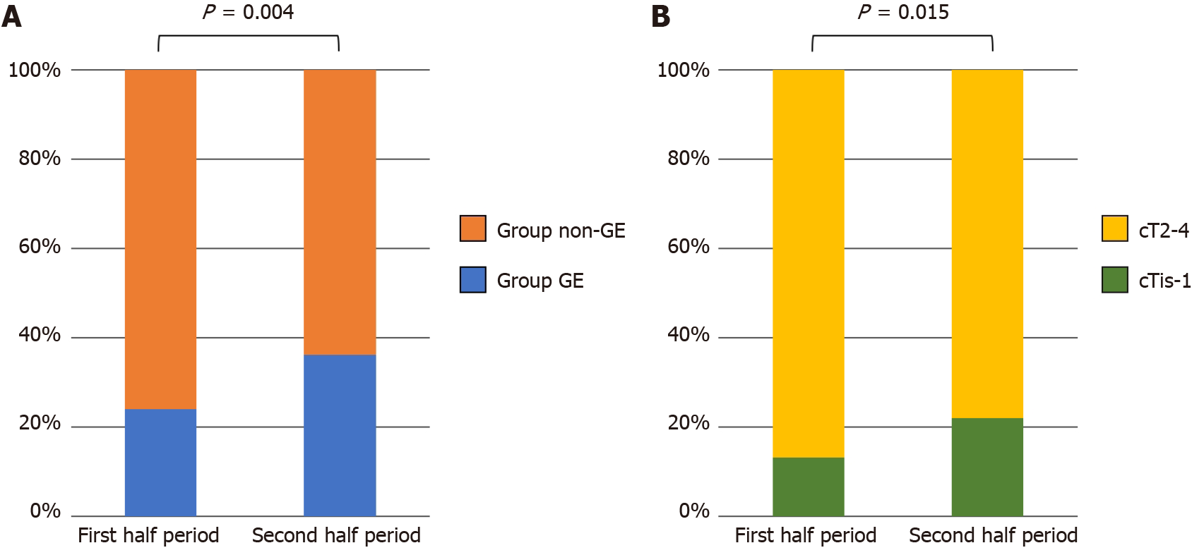
Figure 3 Trends in the detection modality and clinical stage of pharyngeal cancer.
A: A comparison of the proportion of Group gastrointestinal endoscopy between the first and second half periods (2011–2014 and 2015–2018); B: A comparison of the proportion of cTis-1 lesions between first and second half periods (2011–2014 and 2015–2018). Group GE: Group gastrointestinal endoscopy.
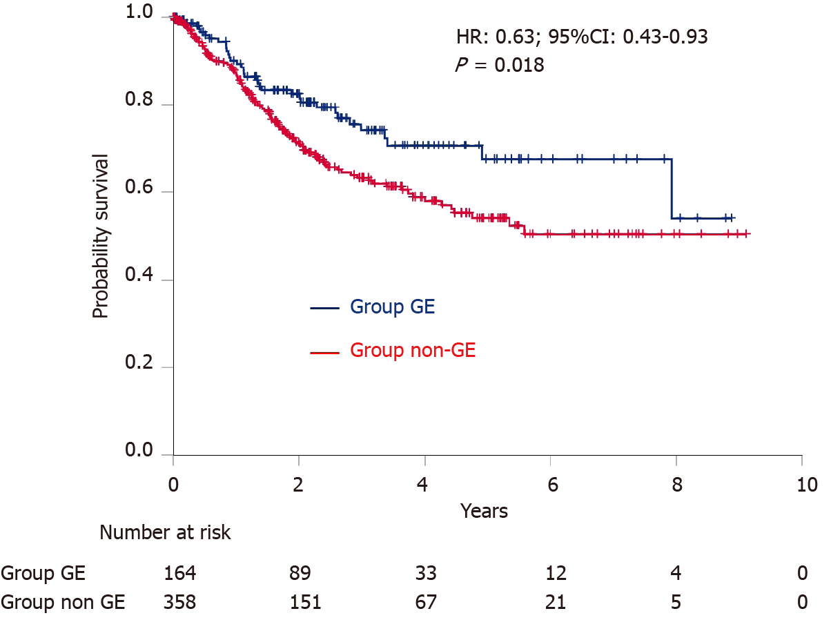
Figure 4 Kaplan–Meier estimates of overall survival.
HR: Hazard ratio; CI: Confidence interval; GE: Gastrointestinal endoscopy.
- Citation: Miyamoto H, Naoe H, Morinaga J, Sakisaka K, Tayama S, Matsuno K, Gushima R, Tateyama M, Shono T, Imuta M, Miyamaru S, Murakami D, Orita Y, Tanaka Y. Clinical impact of gastrointestinal endoscopy on the early detection of pharyngeal squamous cell carcinoma: A retrospective cohort study. World J Gastrointest Endosc 2021; 13(10): 491-501
- URL: https://www.wjgnet.com/1948-5190/full/v13/i10/491.htm
- DOI: https://dx.doi.org/10.4253/wjge.v13.i10.491


