Copyright
©The Author(s) 2020.
World J Gastrointest Endosc. Dec 16, 2020; 12(12): 555-559
Published online Dec 16, 2020. doi: 10.4253/wjge.v12.i12.555
Published online Dec 16, 2020. doi: 10.4253/wjge.v12.i12.555
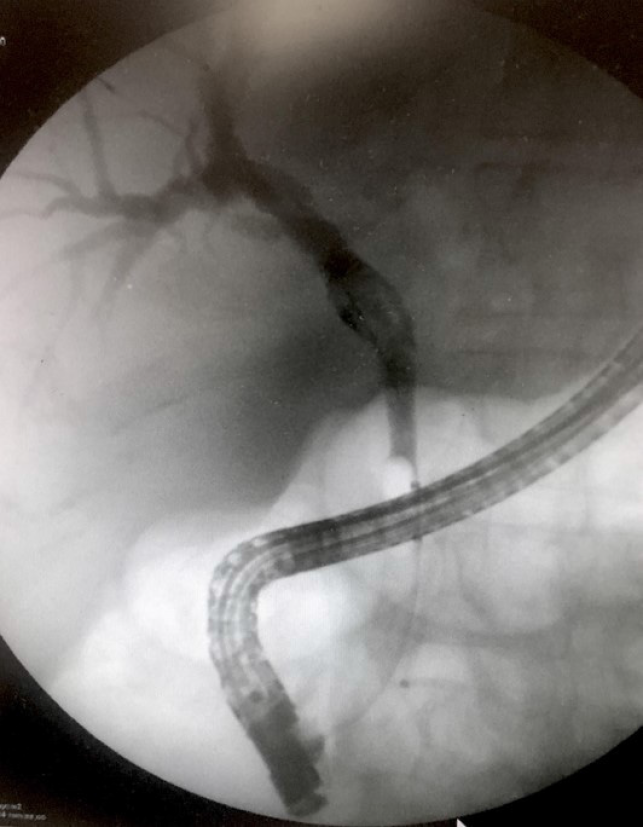
Figure 1 Cholangiography findings during endoscopic retrograde cholangio-pancreatography of the intraductal papillary neoplasm of the bile duct.
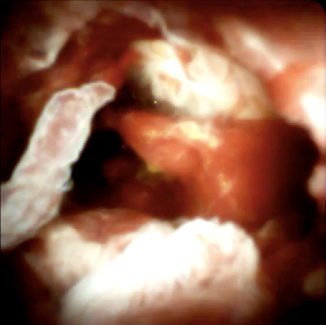
Figure 2 Image of the intraductal papillary neoplasm of the bile duct visualized using the SpyGlass DS II technology.
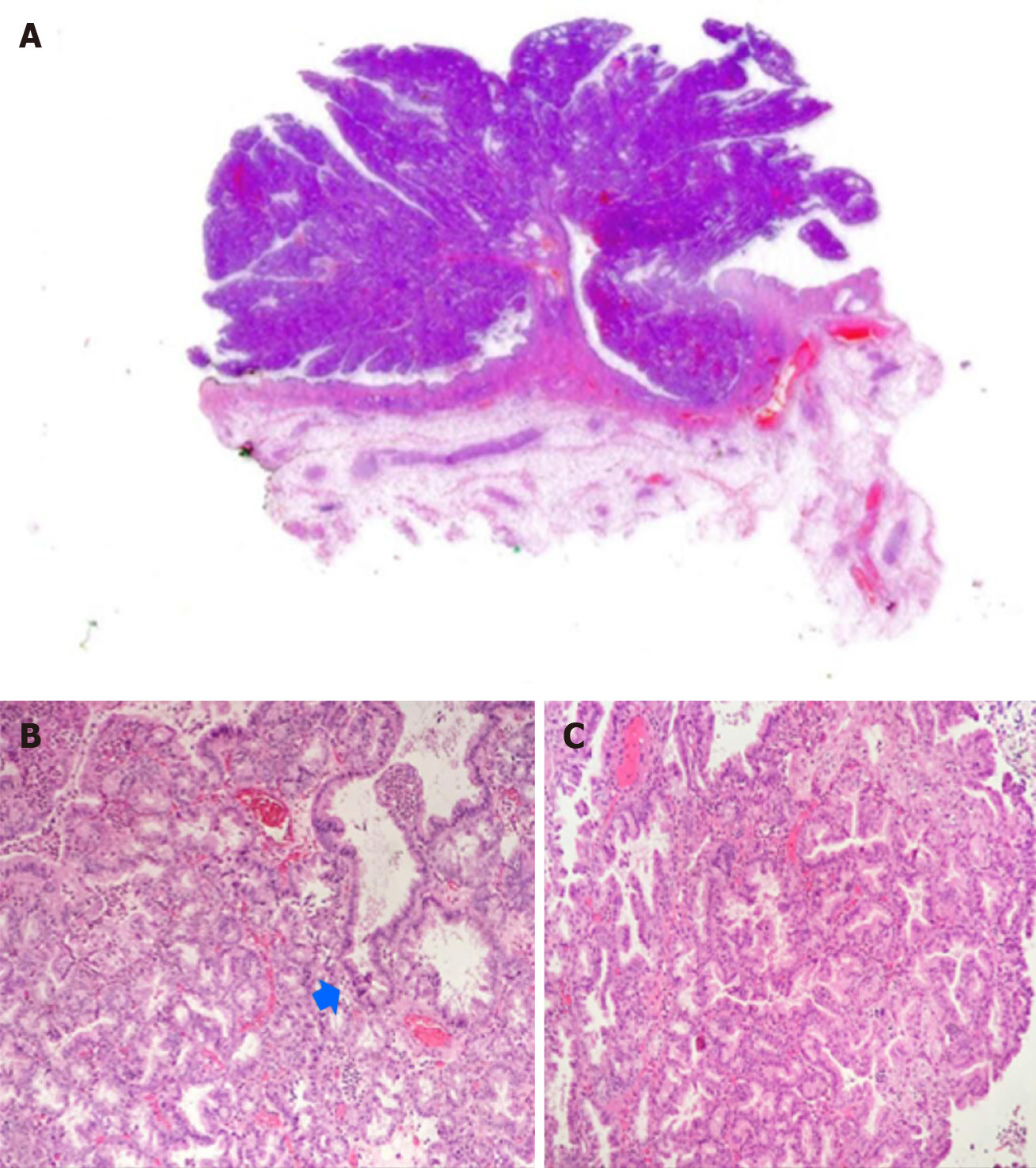
Figure 3 Hematoxylin and eosin staining results.
A: Whole section of the intraductal papillary neoplasm of the bile duct with villous silhouette and pseudo-infiltration of the stroma - arrow (1.25 ×); B and C: Foci of marked cytological atypia in papillomatous background architecture (20 ×).
- Citation: Cocca S, Grande G, Reggiani Bonetti L, Magistri P, Di Sandro S, Di Benedetto F, Conigliaro R, Bertani H. Common bile duct lesions - how cholangioscopy helps rule out intraductal papillary neoplasms of the bile duct: A case report. World J Gastrointest Endosc 2020; 12(12): 555-559
- URL: https://www.wjgnet.com/1948-5190/full/v12/i12/555.htm
- DOI: https://dx.doi.org/10.4253/wjge.v12.i12.555


