Copyright
©The Author(s) 2018.
World J Gastrointest Endosc. Nov 16, 2018; 10(11): 326-339
Published online Nov 16, 2018. doi: 10.4253/wjge.v10.i11.326
Published online Nov 16, 2018. doi: 10.4253/wjge.v10.i11.326
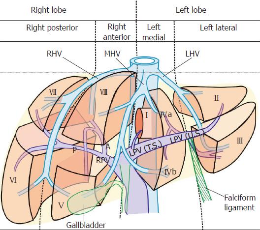
Figure 1 Three vertical planes and a transverse plane divide the liver into four sectors and eight segments.
The vertical planes divide the liver into four sectors. The transverse plane divides the liver into superior and inferior segments. LHV: Left hepatic vein; MHV: Middle hepatic vein; RHV: Right hepatic vein; LPV: Left branch of the portal vein; RPV: Right branch of the portal vein.
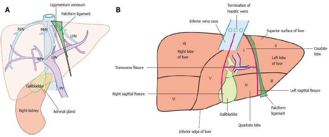
Figure 2 The home bases of imaging and visceral surface of liver.
A: The endoscopic ultrasonography home bases of imaging for liver segments include the inferior vena cava (IVC) during its course behind the liver up to the right atrium, three hepatic veins in portal fissures and their joining point into the IVC, the portal vein and its branches in transverse fissure, the right kidney, the ligamentum teres, the ligamentum venosum, and the gallbladder; B: The caudate lobe lies between the stomach and IVC. The hepatic veins join the IVC. The ligamentum venosum and ligamentum teres are attached to the upper and lower border of the left branch of the portal at the angulation between the transverse and umbilical part. The visceral surface of the liver comes in contact with all segments except segment VIII. PV: Portal vein; LHV: Left hepatic vein; MHV: Middle hepatic vein; RHV: Right hepatic vein; LPV: Left branch of the portal vein; RPV: Right branch of the portal vein.
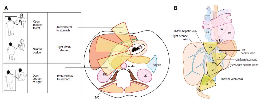
Figure 3 Different operator positions during imaging and three stations of imaging.
A: Rotation is the key movement and should be done in a straight position to transfer the effect of rotation to the tip of the ultrasonic transducer. During imaging from the stomach, the open position to left starts imaging structures placed dorsal to the probe in the esophagus and stomach and primarily screens the left lobe of the liver. The neutral position screens the structures near the liver hilum and the open position to right screens the right lobe of liver; B: The three stations of imaging for liver segments: (1) abdominal part of esophagus (1e) and stomach (1s); (2) duodenal bulb; and (3) second part of the duodenum. IVC: Inferior vena cava; RK: Right kidney; LK: Left kidney; RA: Right atrium; LA: Left atrium; RV: Right ventricle; LV: Left ventricle.
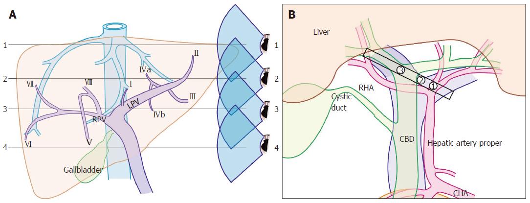
Figure 4 Hepatic vein tributaries with portal vein branches and hilar structures during rotation from left to right edge of transverse fissure.
A: The imaging of hepatic vein tributaries and portal vein branches as the home bases during imaging from the abdominal part of the esophagus and stomach; B: The hilar structures with the divisions during a rotation from the left edge of the transverse fissure to the right edge. (1) hepatic artery into two branches; (2) the union of the right and left branch of the portal vein; (3) the division of the common hepatic duct into right and left branches. LPV: Left branch of the portal vein; RPV: Right branch of the portal vein; CHA: Common hepatic artery; RHA: Right hepatic artery; CBD: Common bile duct.
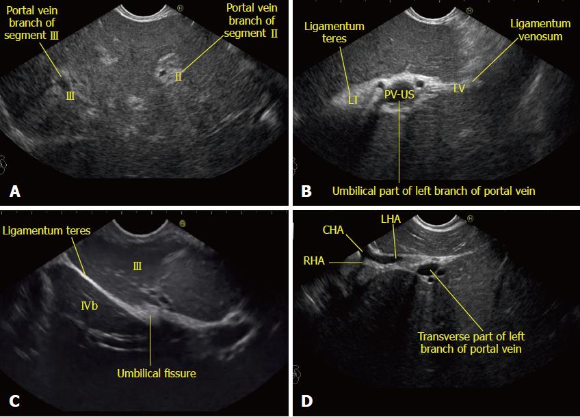
Figure 5 Imaging of liver segments from station 1.
A: Imaging from the abdominal part of esophagus showing segments II and III portal vein branches; B: The fisheye appearance of the umbilical part of the left branch of the portal vein as seen from the abdominal part of esophagus; C: Imaging from the visceral surface of the liver showing that the ligamentum teres is attached to the lower part of the umbilical vein; D: On clockwise rotation near the left edge of the porta hepatis, the umbilical part enters the transverse fissure. At this point the bifurcation of the common hepatic artery can be seen towards the left edge of the transverse fissure. This image shows the entry of the left branch of the common hepatic artery into the transverse fissure. CHA: Common hepatic artery; RHA: Right hepatic artery; LHA: Left hepatic artery.
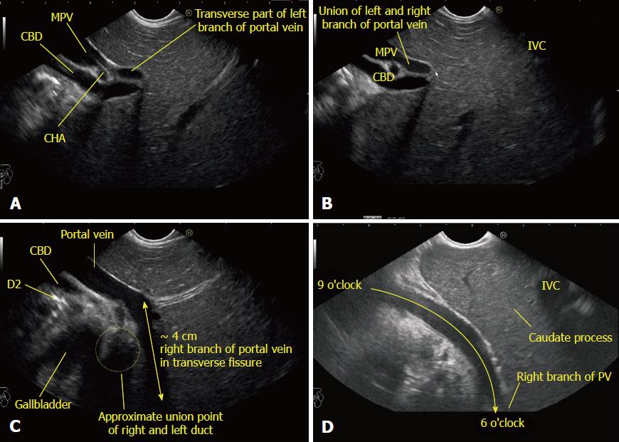
Figure 6 Imaging from neutral position from station 1 showing the tracing of the portal vein and its branches during clockwise rotation from the left to right edge of the transverse fissure.
A: Further clockwise rotation traces the course of the left branch of the portal vein (PV) in the transverse fissure; B: Further rotation shows the union of the right and left branch of the PV; C: The approximate 4 cm breadth of the transverse fissure within which the right branch of the PV joins the left branch; D: The imaging of the right branch of the PV is easy through the caudate process of the liver. With slight up angulation, the right branch of the PV is seen in the transverse fissure going from a 6 o’clock to a 9 o’clock position. CHA: Common hepatic artery; IVC: Inferior vena cava; PV: Portal vein; CBD: Common bile duct; MPV: Main portal vein.
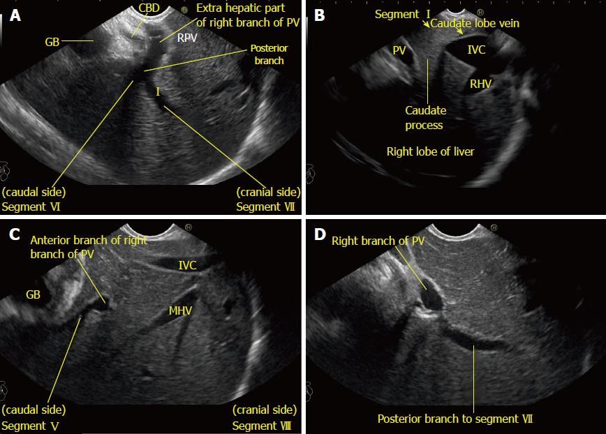
Figure 7 Imaging from station 1 showing the right portal vein and its branches in relation to liver segments.
A: Imaging of the intrahepatic/extrahepatic part of the right branch of the portal vein is seen along with the right lobe of the liver and the hyperechoic diaphragm; B: The inferior vena cava and caudate process provide a good window of imaging for the right lobe of the liver (The caudate lobe is connected with the right lobe of liver through the caudate process). In this location, presence of the inferior vena cava may also provide a satisfactory window of imaging; C: A view of the divisions of the right branch of the portal vein is possible through the caudate process. In this case, the anterior branch is supplying segments V and VIII of the liver; D: The division of segment VI and VII branches is visualized. The upper part of the posterior branch goes towards segment VII. CBD: Common bile duct; GB: Gallbladder; PV: Portal vein; RHV: Right hepatic vein; MHV: Middle hepatic vein; IVC: Inferior vena cava.
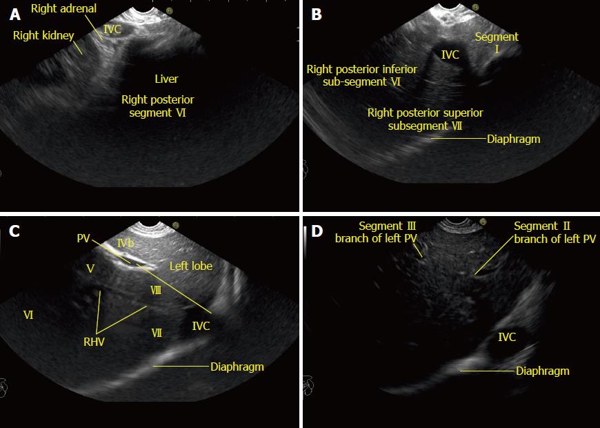
Figure 8 Imaging from the descending duodenum showing the structures visualized during counterclockwise rotation from open position to right.
A: Imaging from the descending duodenum showing the right kidney and inferior vena cava (IVC). The right adrenal gland is seen behind the IVC; B: Imaging from the descending duodenum showing the IVC moving towards the diaphragm. The caudate lobe is seen between the probe and the IVC. The caudate lobe indicates the approximate place of the transverse part of the left branch of the portal vein (PV); C: Imaging from the descending duodenum showing the right hepatic vein. It divides the segments of the right lobe. A line between the cranial end of the IVC and the PV gives approximate locations of the right and left half of the liver; D: The segmental branches (II and III) of the umbilical part of the PV are seen with the IVC at a 4 o’clock position. RHV: Right hepatic vein; IVC: Inferior vena cava; PV: Portal vein.
Figure 9 Imaging from station 1 showing hepatic vein branches and their relationship to liver segments.
A: The middle hepatic vein lies in the course of the Cantlie’s line and separates the left and right lobe of the liver; B: The segmental division is shown for segment I, IVa, IVb, V, and VIII; C: The right hepatic vein separates the right anterior and right posterior sectors; D: The left hepatic vein separates the left medial and left lateral sectors. MHV: Middle hepatic vein; RHV: Right hepatic vein; IVC: Inferior vena cava; PV: Portal vein.
Figure 10 The division of superior and inferior liver segments by the portal vein and its branches.
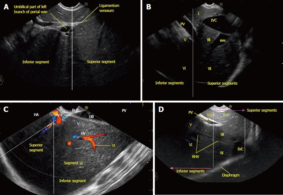
A: Line at the level of the upper border of the umbilical segment of the portal vein dividing the superior and inferior segments; B: The white line divides the superior and inferior segments. This diagram from the esophagus shows the right side of the liver through the caudate lobe of the liver and the inferior vena cava. The presence of hepatoduodenal ligament around the portal vein may not allow a similar quality of visualization of the inferior segments (V and VI); C: This image from station 2 (duodenum bulb) shows the right and middle hepatic veins. The right hepatic vein is parallel to the surface of the gallbladder, and the middle hepatic vein is towards the neck of the gallbladder. Only the right lobe is seen through the gallbladder; D: The right hepatic vein drains segments VI and VII and a variable portion of segments V and VIII. Segment I has direct drainage into the intrahepatic/retrohepatic part of the inferior vena cava. RHV: Right hepatic vein; IVC: Inferior vena cava; PV: Portal vein.
Figure 11 The caudate lobe, its boundaries, and its relationships.
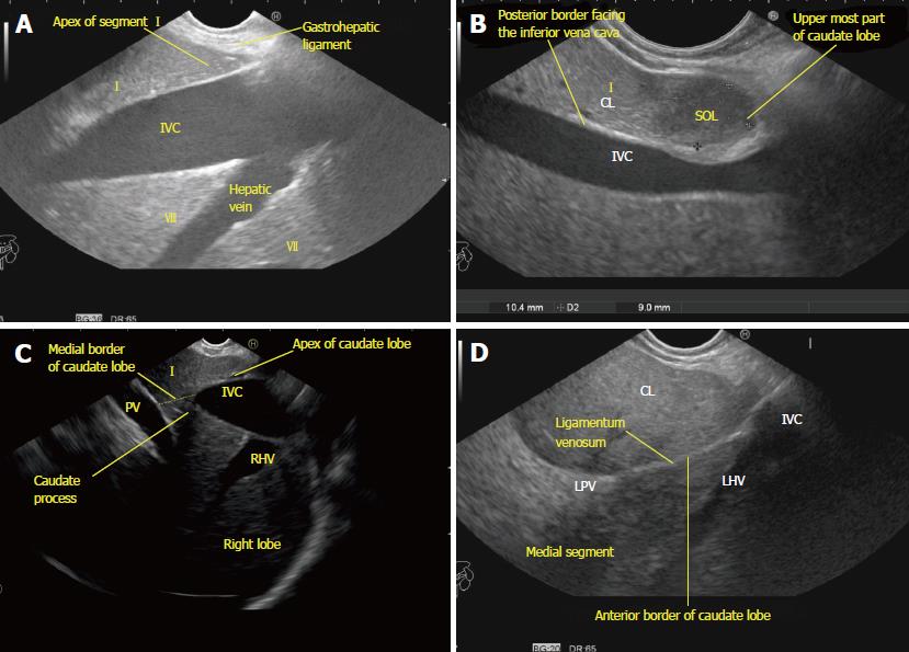
A: The apex of the caudate lobe lies like a wedge near the joining of the hepatic veins. The base faces the inferior vena cava; B: A small metastatic space-occupying lesion is seen near the tip of the caudate lobe of the liver near the diaphragm between the probe and the inferior vena cava. Anterior margin of lesion is limited by the fissure for the ligamentum venosum; C: The continuity of the caudate lobe into the right lobe of the liver via the caudate process; D: The ligamentum venosum proceeds towards the umbilical part of the portal vein and divides the left medial segment from the caudate lobe. The ligamentum venosum is the anterior border of a pyramidal shaped caudate lobe. The attachment of the ligamentum venosum demarcates the lowest limit of the anterior border. SOL: Space-occupying lesion; LHV: Left hepatic vein; LPV: Left branch of the portal vein; RHV: Right hepatic vein; IVC: Inferior vena cava; PV: Portal vein.
Figure 12 The caudate lobe, its boundaries, and its relationships.
A: The exit of the inferior vena cava from the liver demarcates the lowest limit of the posterior border. The right kidney is seen through the inferior vena cava. Segment VI lies above the right kidney; B: The left border of the caudate lobe lies close to superior recess of the lesser sac, which is filled with fluid in this case; C: The vascular supply of the caudate lobe from the right branch of the portal vein. The right and left lobes of the liver lie on either side of the right and left sagittal fissures, and the caudate and quadrate lobes lie posterior and anterior to the transverse fissure. RK: Right kidney; IVC: Inferior vena cava; PV: Portal vein.
Figure 13 The caudate lobe from different stations of imaging.
A: An image of the caudate lobe through the portal vein in front of the inferior vena cava from the duodenal bulb; B: An image of the caudate lobe between the portal vein and the inferior vena cava from the descending duodenum; C: The caudate process acts as a two-way window for imaging of the right and left lobes of the liver in endoscopic ultrasonography. The red circle shows the approximate area of gastric impression on the visceral surface of the left lobe of the liver and shows that the imaging of the right lobe is possible through the caudate lobe and caudate process. The blue circle shows the approximate area of duodenal impression on the visceral surface of the right lobe of the liver and that the imaging of the left lobe is possible through the caudate lobe and caudate process; D: An image of the left lobe of the liver through the caudate lobe from the duodenal bulb. IVC: Inferior vena cava; PV: Portal vein; CBD: Common bile duct; CL: Caudate lobe; LPV: Left branch of the portal vein.
Figure 14 Hepatic veins dividing liver segments.
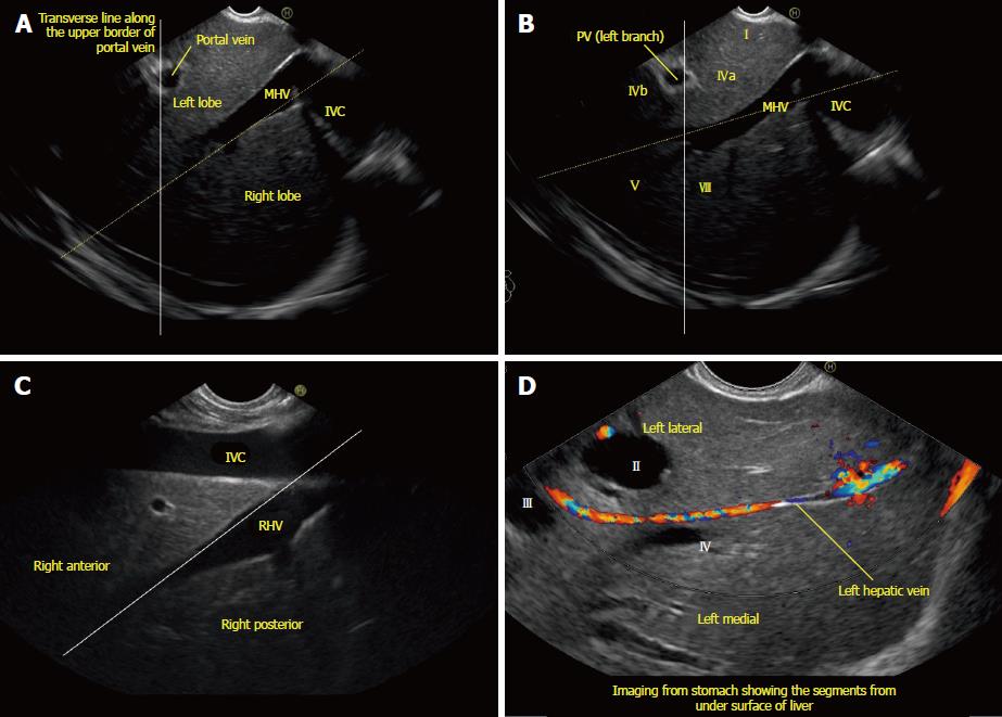
A: Image showing left lobe segments; B: The upper and lower tributaries of the hepatic vein indicate the upper and lower segments of the left lateral and left medial sectors. Segments II, III, IVa, and IVb veins are seen. In this case, the imaging is done from the visceral surface of the liver and from an area close to the antrum and body. Hence, segment IV appears closer to the probe than segment III. It must be clear that while imaging from the lower end of the esophagus that the left lateral segment is closer while during imaging from the visceral surface of the liver that the orientation of a segment may vary depending on the location of probe (for example, near the antrum segment IV is closer than segment III, and near the fundus and proximal body segment III is closer than segment IV). The white line is an extrapolated line that has been drawn in an approximate axis from the upper part of the umbilical part of the left branch of the portal vein; C: A line going through the middle hepatic vein separates the left medial and right anterior sectors; D: A line along the upper part of the transverse fissure (along the upper edge of the portal vein) subdivides the upper and lower segments of the left medial (segments IVa and IVb) and right anterior (segments VIII and V) segments. MHV: Middle hepatic vein; IVC: Inferior vena cava; PV: Portal vein.
Figure 15 Liver sectors and liver segments are visualized.
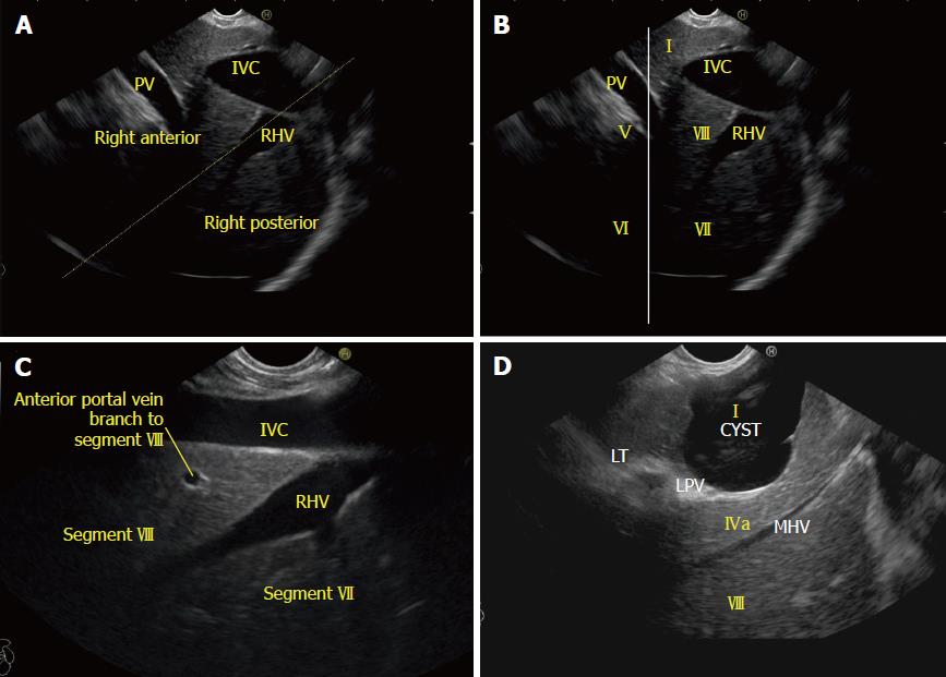
A: The inferior vena cava (IVC) runs parallel to the probe in a long axis. A line along the right hepatic vein divides the liver into anterior and posterior sectors; B: The right lobe of the liver contains segments V to VIII. The segments are seen through the caudate process. The white line is drawn along the upper border of the curving part of the portal vein; C: The right hepatic vein is seen joining the IVC at an angle of around 60°. Segment VII is seen above the hepatic vein and segment VIII is seen between the hepatic vein and the IVC; D: The middle hepatic vein drains segment IV, segment V, and segment VIII. RHV: Right hepatic vein; LPV: Left branch of the portal vein; IVC: Inferior vena cava; PV: Portal vein.
- Citation: Sharma M, Somani P, Rameshbabu CS, Sunkara T, Rai P. Stepwise evaluation of liver sectors and liver segments by endoscopic ultrasound. World J Gastrointest Endosc 2018; 10(11): 326-339
- URL: https://www.wjgnet.com/1948-5190/full/v10/i11/326.htm
- DOI: https://dx.doi.org/10.4253/wjge.v10.i11.326


