Copyright
©The Author(s) 2017.
World J Hepatol. Mar 18, 2017; 9(8): 409-417
Published online Mar 18, 2017. doi: 10.4254/wjh.v9.i8.409
Published online Mar 18, 2017. doi: 10.4254/wjh.v9.i8.409
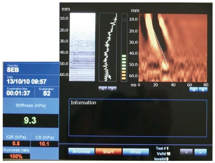
Figure 1 Transient elastography findings for a 10-year-old female suffering from Wilson’s disease.
The patient’s brother had previously developed acute liver failure, which triggered routine monitoring of the patient thereafter. The patient was clinically completely healthy. The transient elastography shows 9.3 kPa, which is above the 6.5 kPa upper limit of normal. Histology findings for the patient showed the liver to be cirrhotic.
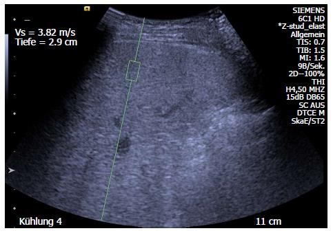
Figure 2 Acoustic radiation force impulse measurement of the liver in a 16-year-old female patient with cystic fibrosis.
Hyperechoic liver parenchyma with irregular liver surface in fibrotic liver parenchyma was revealed. The shear wave velocity was 2.3-3.82 m/s in multiple measurements, significantly above normal values. The same patient had undergone a Fibroscan and the results showed a stiffness of 21.3 ± 2.5 kPa. Six months previously, another Fibroscan had shown a value of 20.4 ± 2.8 kPa.
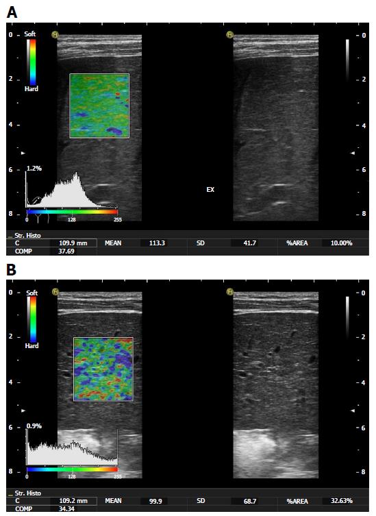
Figure 3 Real-time tissue elastography in a normal and cirrhotic liver.
A: RTE with a normal strain histogram (mean: 113.3 a.u.; %AREA: 10%) in a 8-year-old female patient with cystic fibrosis and nearly normal liver structure; B: RTE with pathological strain histogram in a 6-year-old female patient with tyrosinemia type 1 and liver cirrhosis with small nodules. The mean value was 99.9, and the peak of histogram shifted to the left to lower values of the mean. The percentage of stiffer areas (colour-coded in blue; %AREA) increased up to 23.6%. This histogram is more flattened in comparison to the normal strain histogram. RTE: Real-time tissue elastography.
- Citation: Engelmann G, Quader J, Teufel U, Schenk JP. Limitations and opportunities of non-invasive liver stiffness measurement in children. World J Hepatol 2017; 9(8): 409-417
- URL: https://www.wjgnet.com/1948-5182/full/v9/i8/409.htm
- DOI: https://dx.doi.org/10.4254/wjh.v9.i8.409


