Copyright
©The Author(s) 2016.
World J Hepatol. Jun 18, 2016; 8(17): 731-738
Published online Jun 18, 2016. doi: 10.4254/wjh.v8.i17.731
Published online Jun 18, 2016. doi: 10.4254/wjh.v8.i17.731
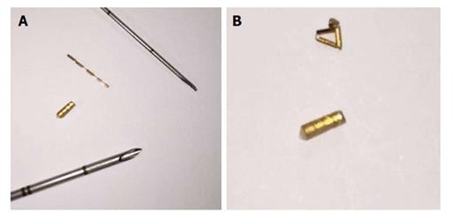
Figure 1 Types of gold fiducial markers.
A: Twenty five gauge and 17 G needle and their gold markers: Grain cylindrical gold marker, 1 mm × 4 mm and flexible wire notched gold marker 0.28 mm × 10 mm, Gold Anchor Marker respectively; B: Grain cylindrical gold marker, 1 mm × 4 mm flexible wire notched gold marker 0.28 mm × 10 mm, Gold Anchor marker after massing.
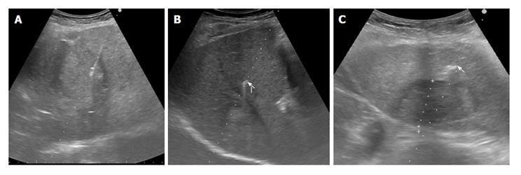
Figure 2 Ultrasonography-guided fiducial placement.
The two different gold markers are sonographically undistinguishable. A: Needle delivering fiducial into a liver mass; B: Hyperechoic flexible wire notched gold marker, 0.28 mm × 10 mm, Gold Anchor marker (arrow) near a liver mass; C: Hyperechoic Grain cylindrical gold marker, 1 mm × 4 mm (arrow) near a liver mass.
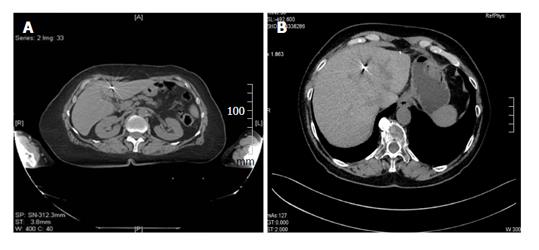
Figure 3 Well visualized fiducial markers.
A: CT displaying of Grain cylindrical gold marker, 1 mm × 4 mm. This marker exhibits good contrast on X-ray images with typical “star effects”; B: CT displaying types of flexible wire notched gold marker, 0.28 mm × 10 mm, Gold Anchor marker. This marker exhibits good contrast on X-ray images demonstrating less artifacts. CT: Computed tomography.
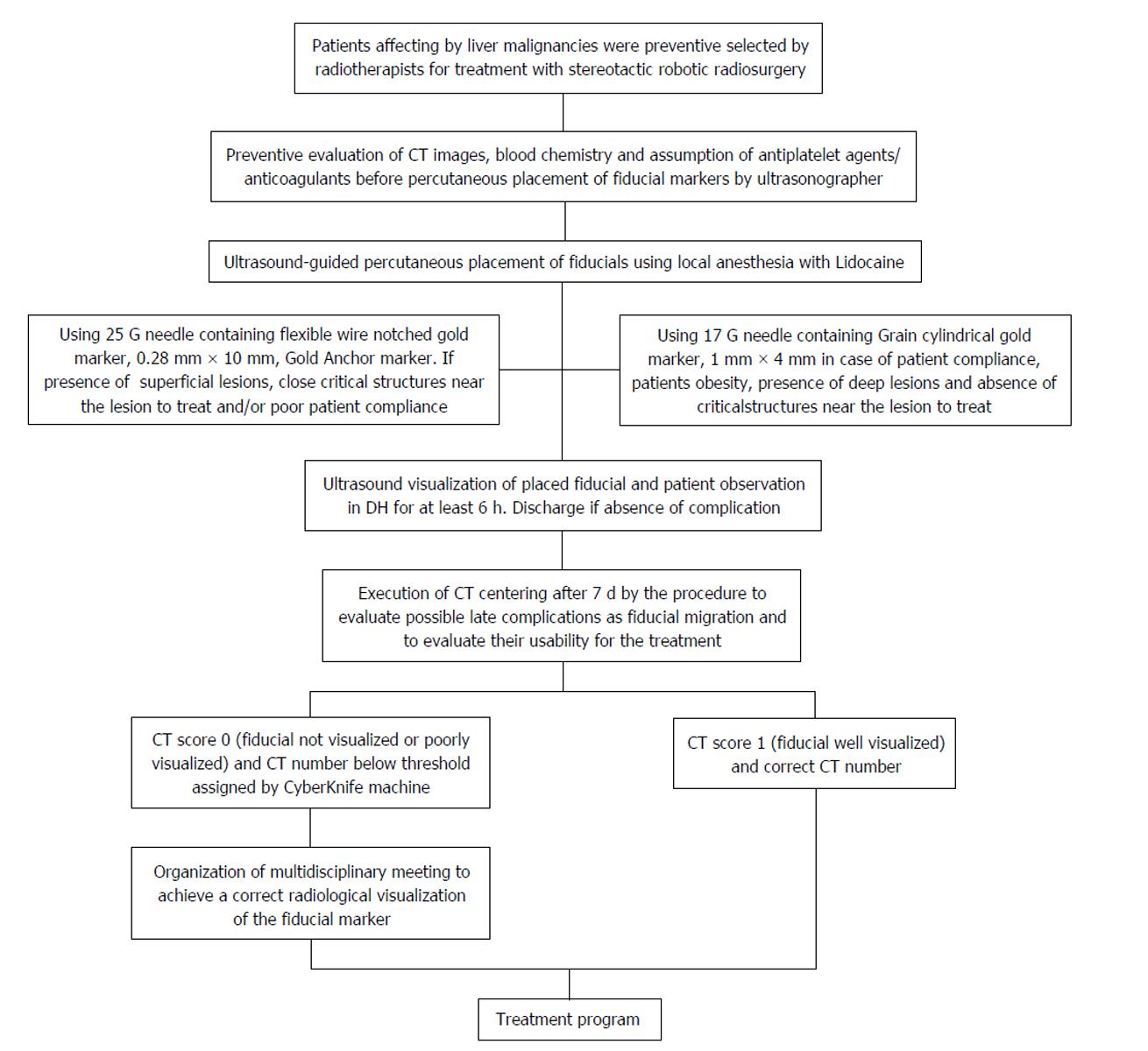
Figure 4 Flow chart of the study conducted tank to a prospective collection of data (compliance, demographic and clinic characteristics of the patients, liver lesions characteristics, type of needle and markers used, ultrasonographic and computed tomography visualization of fiducial markers, usability of the markers and immediate and late complications) and a retrospective statistical analysis.
CT: Computed tomography.
- Citation: Marsico M, Gabbani T, Livi L, Biagini MR, Galli A. Therapeutic usability of two different fiducial gold markers for robotic stereotactic radiosurgery of liver malignancies: A pilot study. World J Hepatol 2016; 8(17): 731-738
- URL: https://www.wjgnet.com/1948-5182/full/v8/i17/731.htm
- DOI: https://dx.doi.org/10.4254/wjh.v8.i17.731


