Copyright
©The Author(s) 2016.
World J Hepatol. Jun 8, 2016; 8(16): 691-702
Published online Jun 8, 2016. doi: 10.4254/wjh.v8.i16.691
Published online Jun 8, 2016. doi: 10.4254/wjh.v8.i16.691
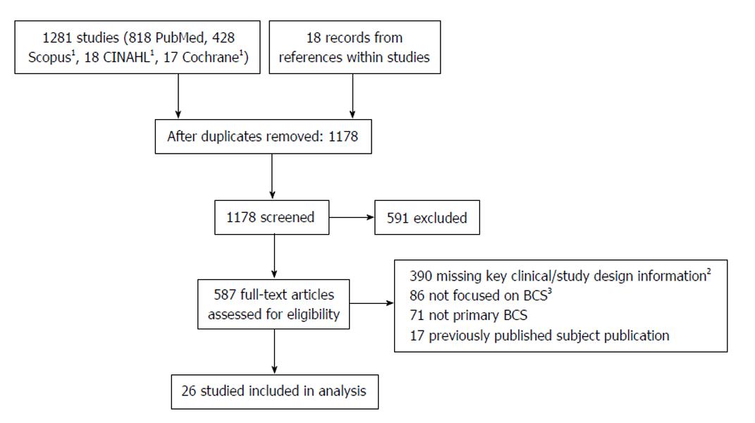
Figure 1 Flow diagram of studies selection.
1Searches conducted with MEDLINE results removed; 2Studies missing key clinical information including clear inclusion and exclusion criteria, clear diagnostic parameters, etc., and studies that investigated subpopulations (e.g., BCS patients requiring liver transplantation, BCS patients without MPN, etc.); 3Studies focused on other categories (e.g., causes of liver failure). BCS: Budd-Chiari syndrome; MPN: Myeloproliferative neoplasms.
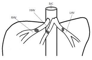
Figure 2 Classical Budd-Chiari syndrome - Occlusions are within the hepatic veins themselves and usually thrombi.
RHV: Right hepatic vein; MHV: Middle hepatic vein; LHV: Left hepatic vein; IVC: Inferior vena cava.
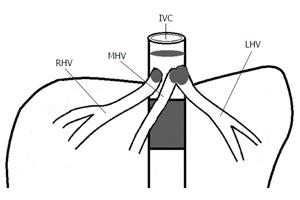
Figure 3 Hepatic vena cava-Budd Chiari syndrome - Occlusions are thin or thick (membranous or segmental) and within the inferior vena cava and occlusion can extend into the hepatic veins and generally involve the ostia to the inferior vena cava.
RHV: Right hepatic vein; MHV: Middle hepatic vein; LHV: Left hepatic vein; IVC: Inferior vena cava.
- Citation: Shin N, Kim YH, Xu H, Shi HB, Zhang QQ, Colon Pons JP, Kim D, Xu Y, Wu FY, Han S, Lee BB, Li LS. Redefining Budd-Chiari syndrome: A systematic review. World J Hepatol 2016; 8(16): 691-702
- URL: https://www.wjgnet.com/1948-5182/full/v8/i16/691.htm
- DOI: https://dx.doi.org/10.4254/wjh.v8.i16.691


