Copyright
©2014 Baishideng Publishing Group Inc.
World J Hepatol. Jul 27, 2014; 6(7): 532-537
Published online Jul 27, 2014. doi: 10.4254/wjh.v6.i7.532
Published online Jul 27, 2014. doi: 10.4254/wjh.v6.i7.532
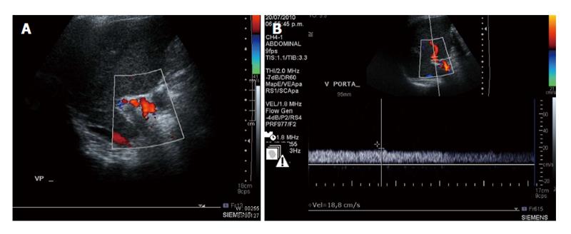
Figure 1 Doppler ultrasound.
A: Liver Doppler ultrasound. The image shows the thrombus in the portal vein; B: Doppler ultrasound, performed 4 mo after discharge, revealed that the portal vein thrombi had disappeared and a smooth bloodstream was observed in the portal vein.
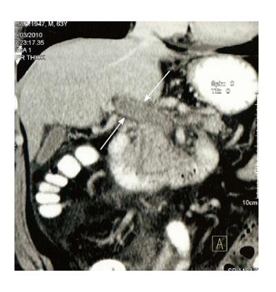
Figure 2 Coronal reconstruction of contrast-enhanced computed tomography image with arrows indicating portal venous thrombosis and evidence of cavernous transformation.
- Citation: Rodríguez-Leal GA, Morán S, Corona-Cedillo R, Brom-Valladares R. Portal vein thrombosis with protein C-S deficiency in a non-cirrhotic patient. World J Hepatol 2014; 6(7): 532-537
- URL: https://www.wjgnet.com/1948-5182/full/v6/i7/532.htm
- DOI: https://dx.doi.org/10.4254/wjh.v6.i7.532


