Copyright
©The Author(s) 2025.
World J Hepatol. Jul 27, 2025; 17(7): 107378
Published online Jul 27, 2025. doi: 10.4254/wjh.v17.i7.107378
Published online Jul 27, 2025. doi: 10.4254/wjh.v17.i7.107378
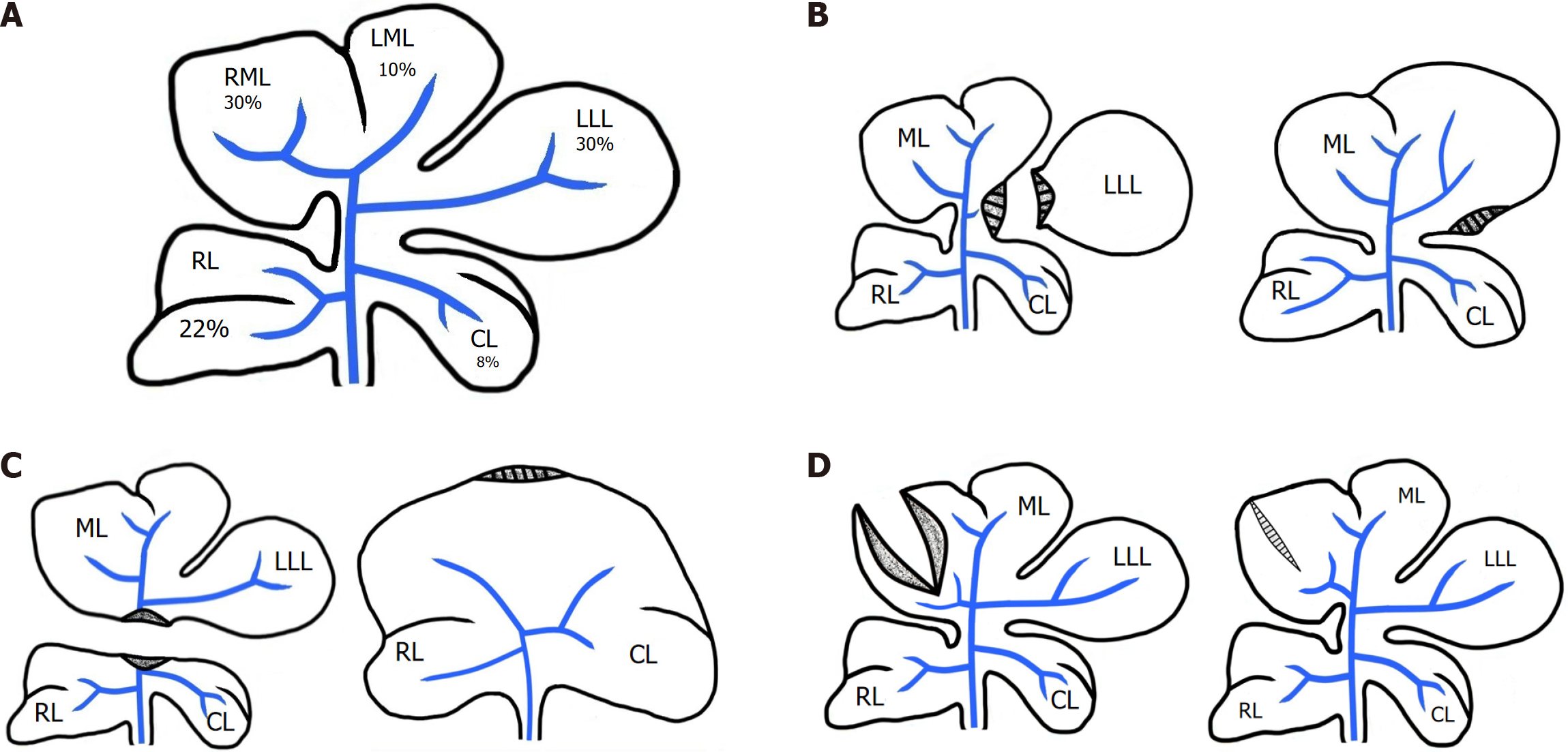
Figure 1 Schematic representation of rat liver lobes resection variations and Subsequent processes in the liver.
A: Normal rat liver; B: Resection of 30% of the liver mass – the Left Lateral Lobe (LLL) – followed by hypertrophic regeneration; C: Resection of 70% of the liver mass – the Right Median Lobe (RML), Left Median Lobe (LML), and LLL – followed by hyperplastic regeneration; D: Dissection of the median lobe (ML) and the subsequent healing and scar formation; RL: Right lateral lobe; CL: Caudate lobe; ML: Median lobe; LLL: Left lateral lobe; RML: Right Median Lobe, LML: Left Median Lobe.
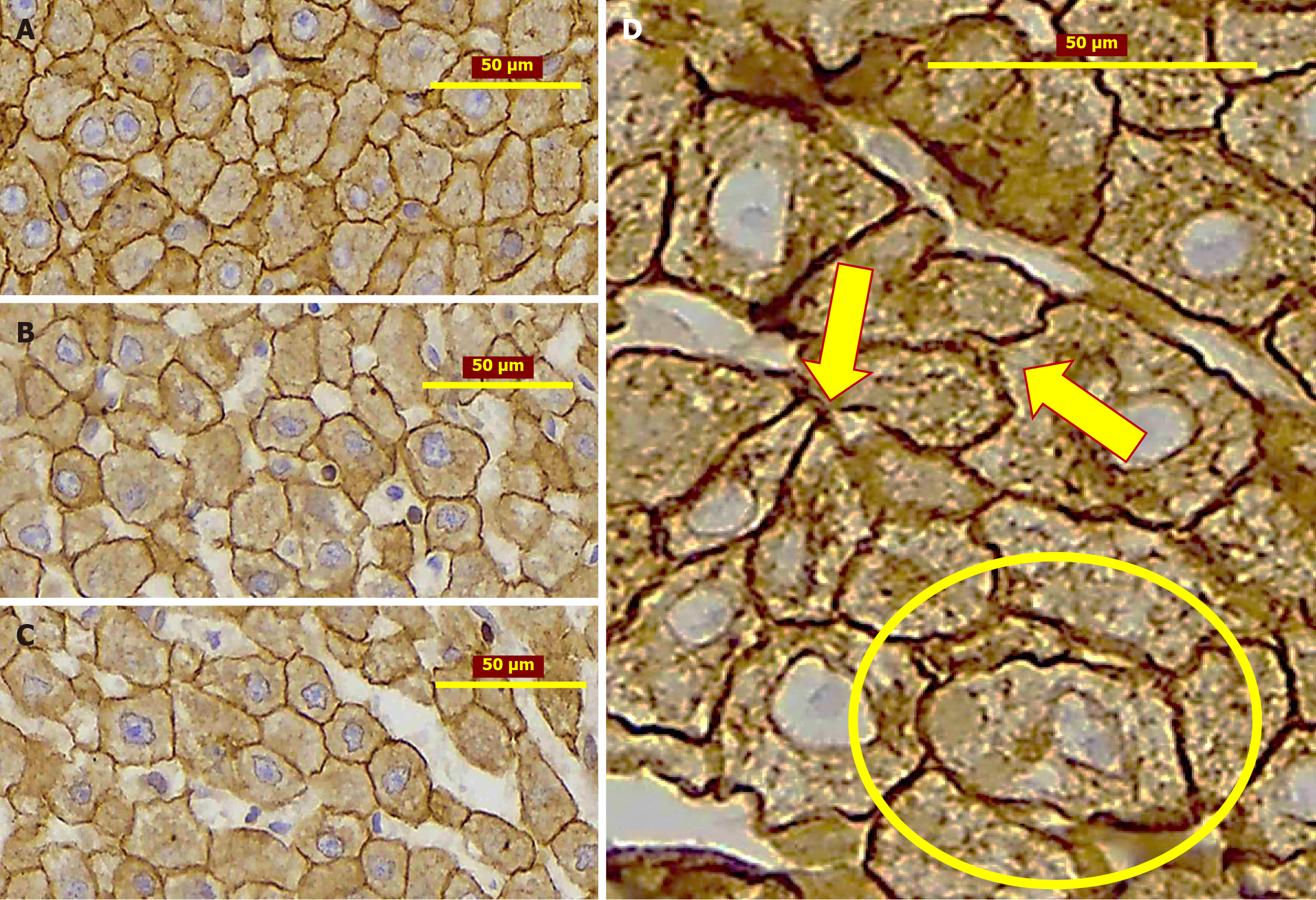
Figure 2 Remodeling of hepatocytes during liver regeneration.
A: Polymorphic Hepatocytes, after 24 hours from partial hepatectomy (PH); B: Polymorphic Hepatocytes after 1 week from PH; C: Polymorphic Hepatocytes after 2 weeks from PH; D: Polymorphic Hepatocytes with protrusions (arrow) and zig-zag formed membrane (circle). IHC stain, CK8. Male Wistar rats.
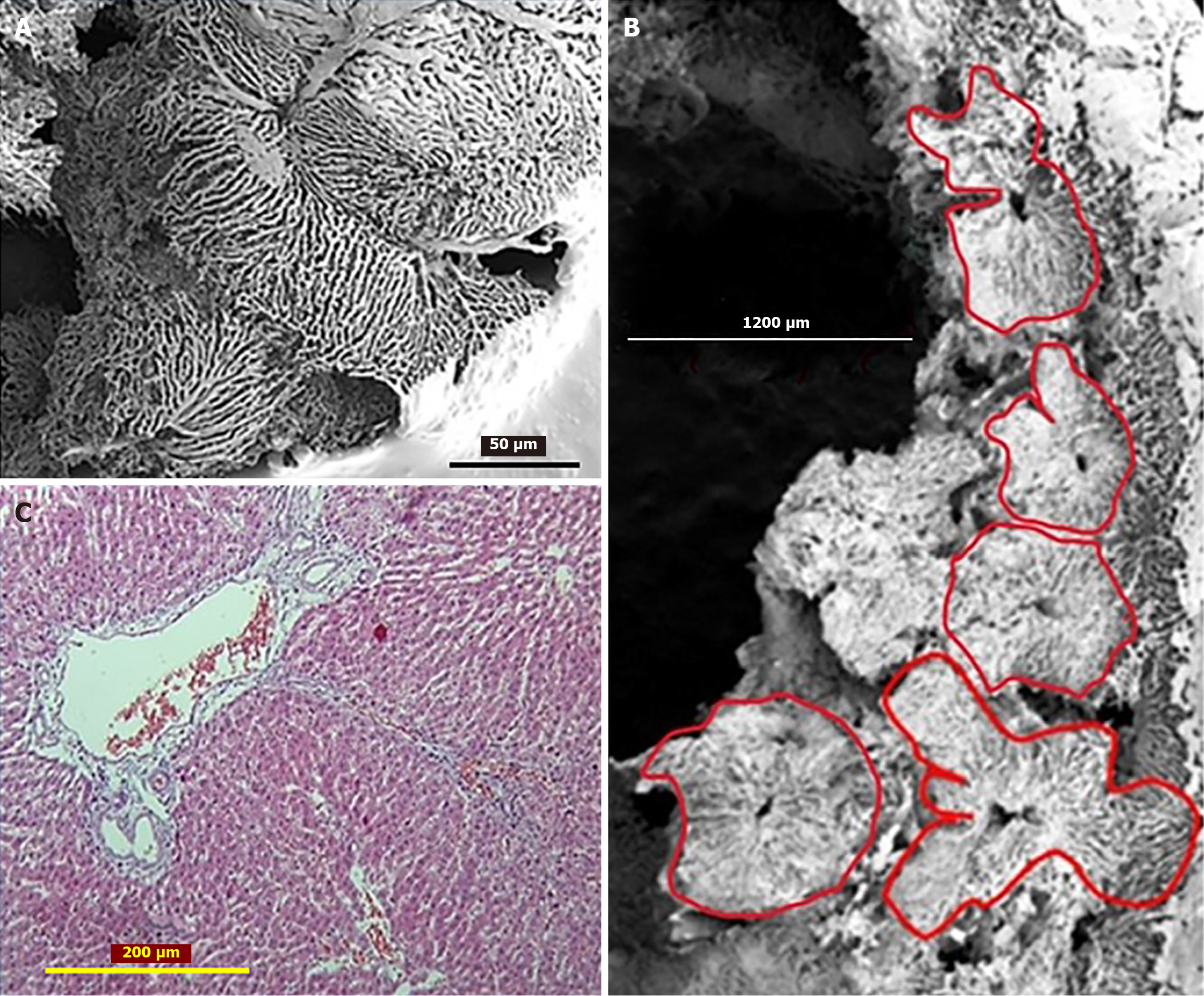
Figure 3 Lobular remodeling following partial hepatectomy.
A and B: The corrosion casts of the sinusoidal network forming the lobules of different sizes and shapes. SEM of Corrosion casts; C: The boundaries between the adjacent lobules are widened and filled by connective tissue fibers. 6 months after partial hepatectomy. Masson’s Trichrome; Male Wistar rats.
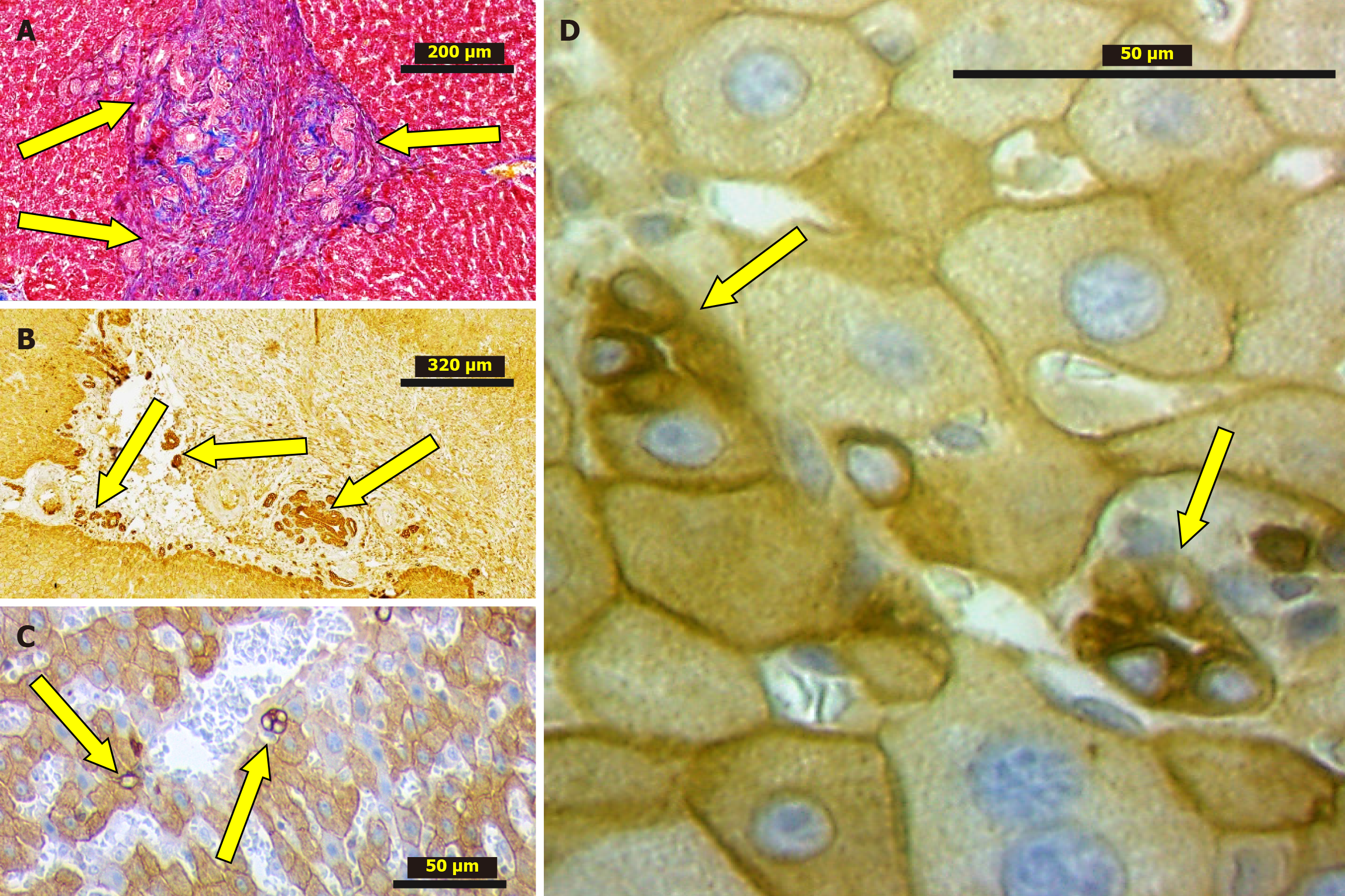
Figure 4 Ductular reaction (arrows).
A: The granulation tissue developed at the area of liver lobes adhesion after 2 weeks from partial hepatectomy (PH). Masson’s Trichrome; B: The granulation tissue developed at the area cut edge necrosis 2 weeks from PH. IHC stain, CK8; C and D: Hepatic lobules after 6 months from PH. IHC stain, CK8. Male Wistar rats.
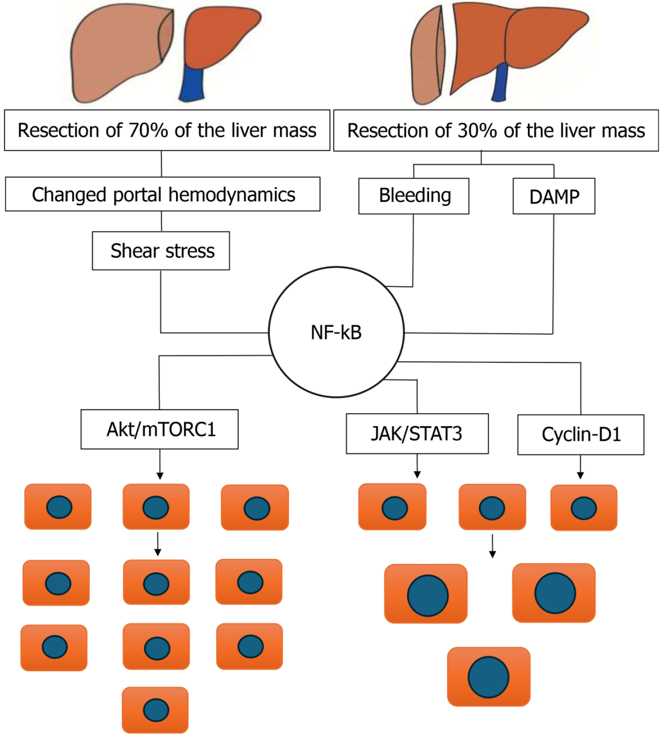
Figure 5 The dual possibility for nuclear factor-kappa B to be involved in hepatocytes hypertrophy as well as hepatocytes hyperplasia during liver regeneration.
DAMP: Damage-associated molecular patterns; NF-kB: Nuclear factor kappa-light-chain-enhancer of activated B cells; Akt/mTORC1: Akt/mammalian target of rapamycin complex 1; JAK/STAT3: Janus kinase/ signal transducer and activator of transcription 3.
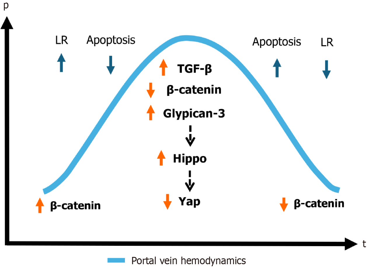
Figure 6 Termination of liver regeneration.
p: Portal pressure; t: Time passed from 70% partial hepatectomy; LR: Liver regeneration; TGF-β: Transforming growth factor beta; YAP: Yes-associated protein.
- Citation: Korchilava B, Khachidze T, Megrelishvili N, Svanadze L, Kakabadze M, Tsomaia K, Jintcharadze M, Kordzaia D. Liver regeneration after partial hepatectomy: Triggers and mechanisms. World J Hepatol 2025; 17(7): 107378
- URL: https://www.wjgnet.com/1948-5182/full/v17/i7/107378.htm
- DOI: https://dx.doi.org/10.4254/wjh.v17.i7.107378


