Copyright
©The Author(s) 2025.
World J Hepatol. Jan 27, 2025; 17(1): 100392
Published online Jan 27, 2025. doi: 10.4254/wjh.v17.i1.100392
Published online Jan 27, 2025. doi: 10.4254/wjh.v17.i1.100392
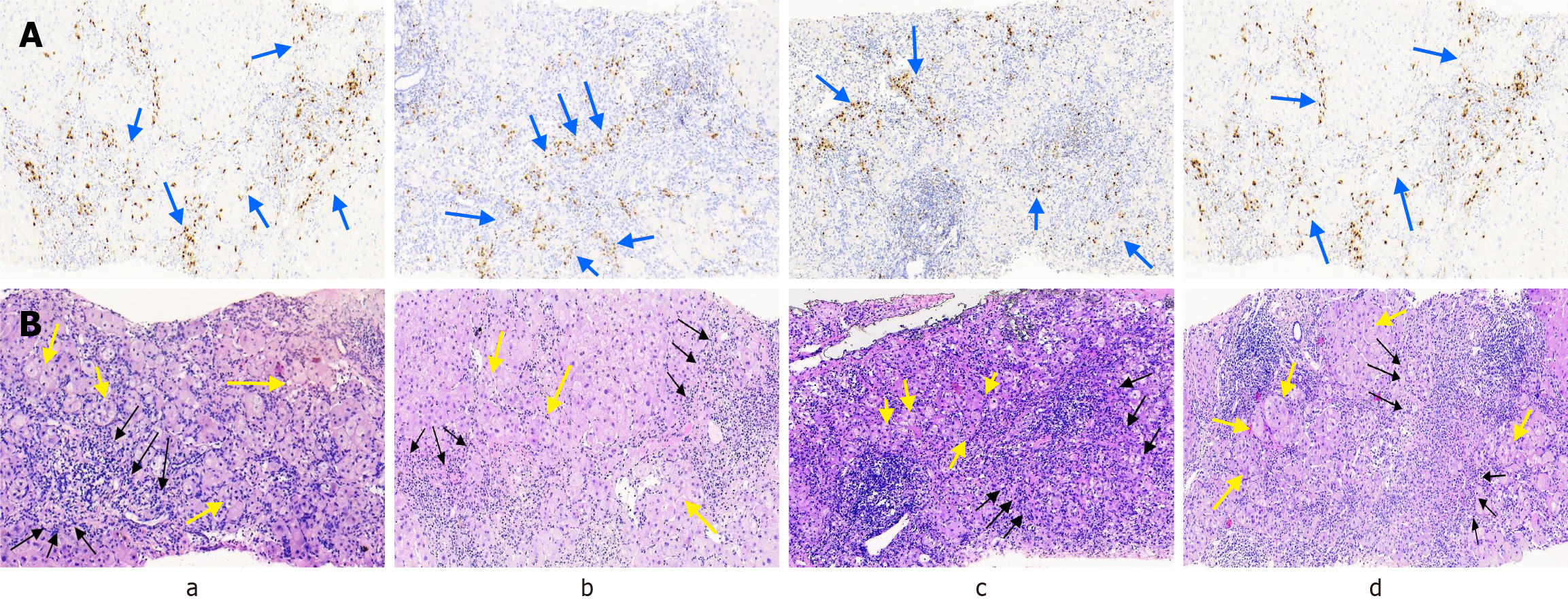
Figure 1 Histological characteristics of liver in patients 8, 9, 10, and 12 (a, b, c, and d).
A: MUM-1 immunohistochemistry: A large number of plasma cells infiltrated in the hepatic lobules, and plasma cells concentrated in the sink area and interface (blue arrow); B: Hematoxylin and eosin staining: Rosettes (yellow arrow) extensive inflammatory cell infiltration in the hepatic lobules, moderate to severe interfacial hepatitis (black arrow).
- Citation: Jiang ML, Xu F, Li JL, Luo JY, Hu JL, Zeng XQ. Clinical features of abnormal α-fetoprotein in 15 patients with chronic viral hepatitis B after treatment with antiviral drugs. World J Hepatol 2025; 17(1): 100392
- URL: https://www.wjgnet.com/1948-5182/full/v17/i1/100392.htm
- DOI: https://dx.doi.org/10.4254/wjh.v17.i1.100392


