Copyright
©The Author(s) 2024.
World J Hepatol. Nov 27, 2024; 16(11): 1348-1355
Published online Nov 27, 2024. doi: 10.4254/wjh.v16.i11.1348
Published online Nov 27, 2024. doi: 10.4254/wjh.v16.i11.1348
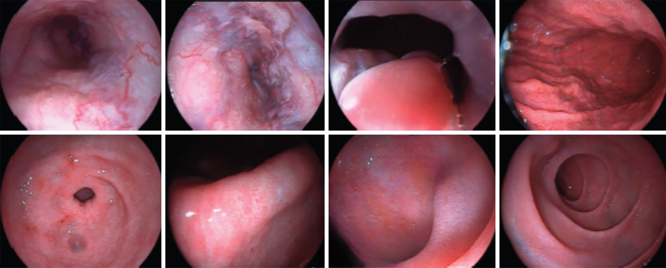
Figure 1 Severe esophageal and gastric varices (positive red sign) and portal hypertensive gastropathy with erosion.
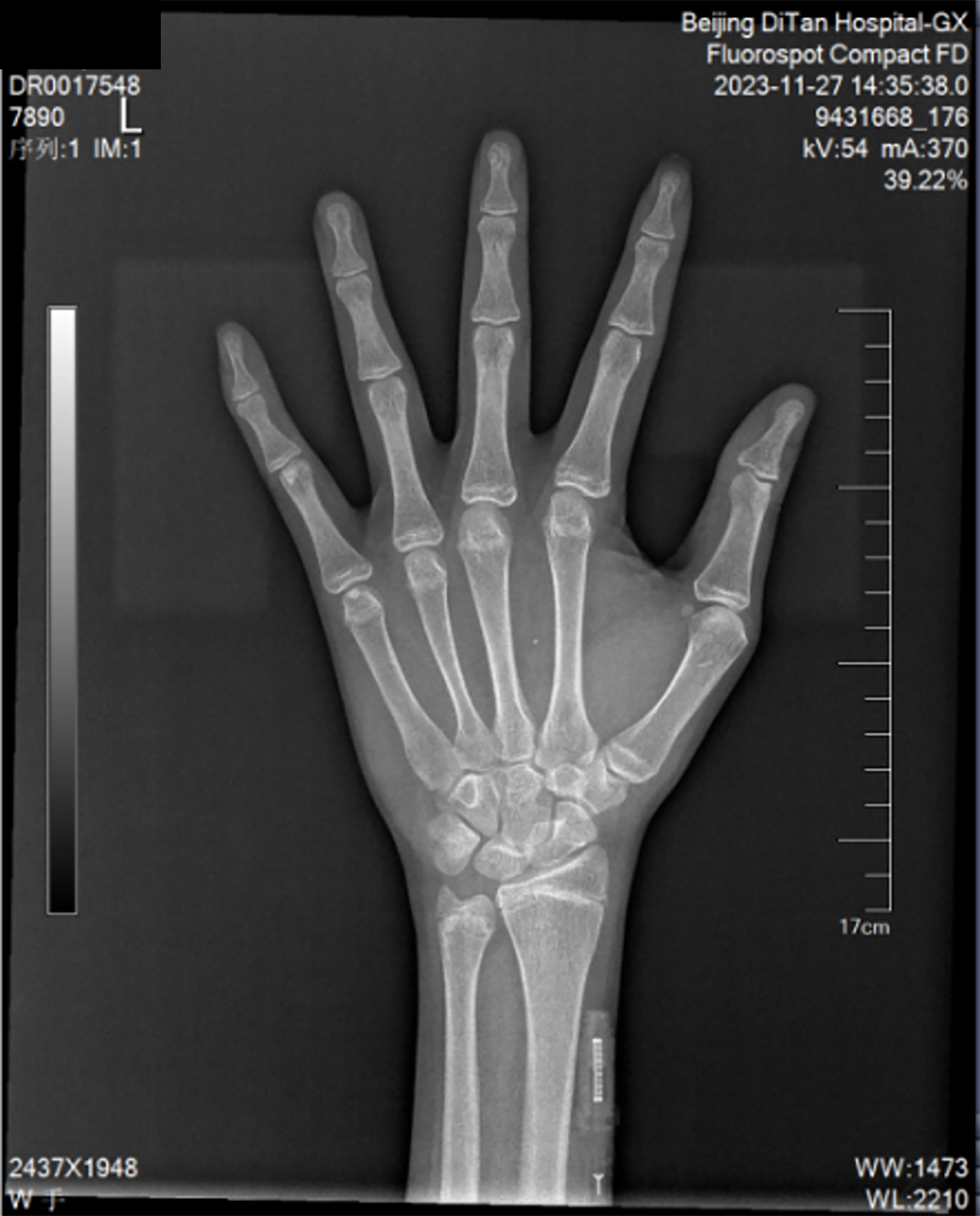
Figure 2 Bone age of the left hand (the patient is right-handed) showed that the bone cortex was thinner, the trabeculae were sparse, and the metacarpal, phalangeal, ulnar, and radial epiphysis had just been closed.
Figure 3 Abdominal enhanced computed tomography plus three-dimensional reconstruction of the portal vein.
The shape of the liver was irregular, the surface of the liver was not smooth, the proportion of each lobe was abnormal, the spleen was obviously enlarged, the esophageal and gastric fundus varices were mild, and the splenic vein was obviously widened. A: The arterial phase demonstrated disproportionate lobar distribution of the liver, widened fissures, and nodular changes in the hepatic parenchyma, with no abnormal enhancement detected in the liver on contrast-enhanced imaging; B: Coronal view revealed significant splenomegaly; C: Coronal view showed marked dilation of the splenic vein, with the splenic vein diameter exceeding that of the portal vein; D: Three-dimensional imaging highlighted prominent dilation of the splenic vein.
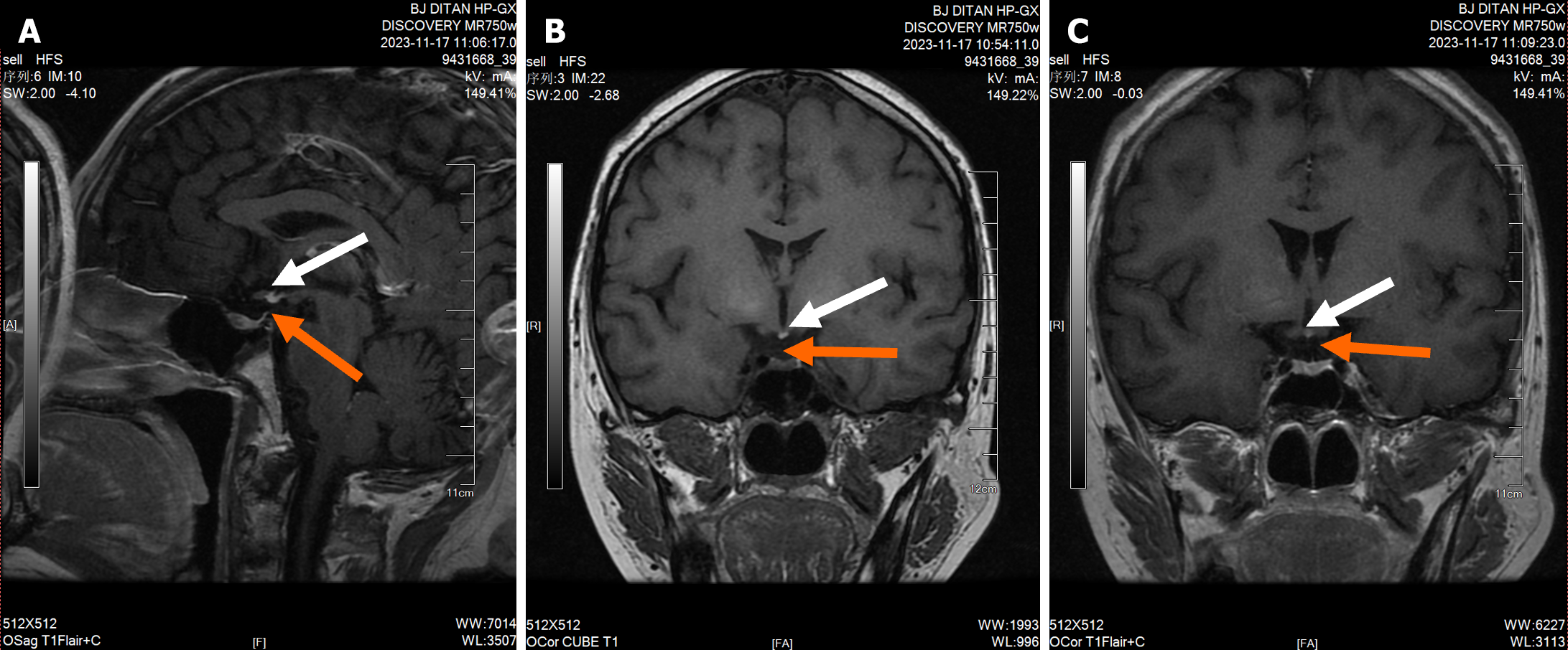
Figure 4 Cranial magnetic resonance imaging results.
Absence of pituitary stalk and ectopic posterior pituitary. The red arrow shows the position of the pituitary stalk, and the white arrow indicates the ectopic posterior pituitary. A: The mid-sagittal plane showed a normal pituitary volume, absence of visualization of the pituitary stalk, and loss of the normal hyperintensity of the posterior pituitary lobe. An ectopic high signal was observed at the infundibular recess; B and C: Coronal planes demonstrated the absence of visualization of the pituitary stalk and loss of the normal hyperintensity of the posterior pituitary lobe, with an ectopic high signal at the infundibular recess.
- Citation: Chang M, Wang SY, Zhang ZY, Hao HX, Li XG, Li JJ, Xie Y, Li MH. Pituitary stalk interruption syndrome complicated with liver cirrhosis: A case report. World J Hepatol 2024; 16(11): 1348-1355
- URL: https://www.wjgnet.com/1948-5182/full/v16/i11/1348.htm
- DOI: https://dx.doi.org/10.4254/wjh.v16.i11.1348


