Copyright
©The Author(s) 2021.
World J Hepatol. Jun 27, 2021; 13(6): 709-716
Published online Jun 27, 2021. doi: 10.4254/wjh.v13.i6.709
Published online Jun 27, 2021. doi: 10.4254/wjh.v13.i6.709
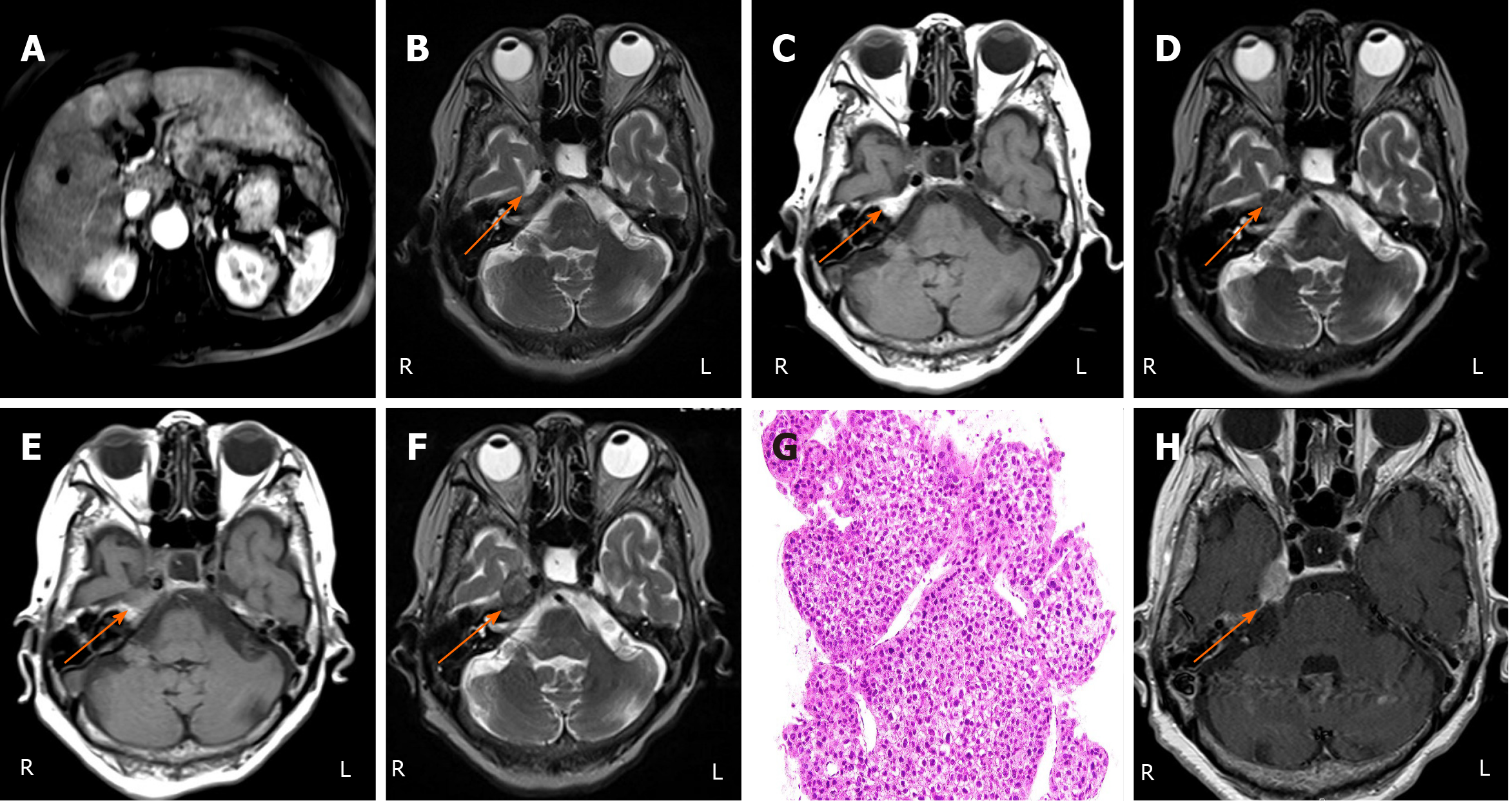
Figure 1 Imaging findings and histopathological findings.
A: Ethoxybenzyl magnetic resonance imaging (MRI), hypervascular hepatocellular carcinoma (HCC) in the right and left lobes; B: Brain MRI [T2-weighted image (T2WI)], no findings in the cavernous sinus or Meckel’s cave; C: Brain MRI [T1-weighted image (T1WI)], intact findings of bone marrow in the petrous bone; D: Brain MRI (T2WI), low intensity mass in the right Meckel’s cave (arrow); E: Brain MRI (T1WI), loss of normal fatty bone marrow signal intensity in the right petrous bone (or apex); F: Brain MRI (T2WI), low intensity mass around the right cavernous node, the right Meckel's cave, and the right petrous bone on T2WI; G: Histopathological finding (hematoxylin and eosin staining), moderately differentiated HCC; H: Contrast enhanced MRI, well-defined mass with abnormal enhancement in the right cavernous sinus, and the right Meckel’s cave (arrow). L: left; R: Right.
- Citation: Kim SK, Fujii T, Komaki R, Kobayashi H, Okuda T, Fujii Y, Hayakumo T, Yuasa K, Takami M, Ohtani A, Saijo Y, Koma YI, Kim SR. Distant metastasis of hepatocellular carcinoma to Meckel’s cave and cranial nerves: A case report and review of literature. World J Hepatol 2021; 13(6): 709-716
- URL: https://www.wjgnet.com/1948-5182/full/v13/i6/709.htm
- DOI: https://dx.doi.org/10.4254/wjh.v13.i6.709


