Copyright
©The Author(s) 2020.
World J Hepatol. Mar 27, 2020; 12(3): 108-115
Published online Mar 27, 2020. doi: 10.4254/wjh.v12.i3.108
Published online Mar 27, 2020. doi: 10.4254/wjh.v12.i3.108
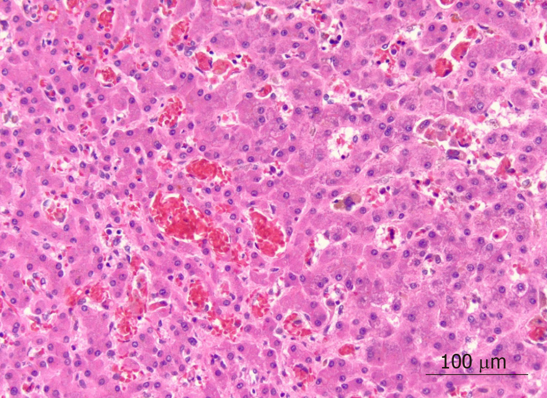
Figure 1 Liver parenchyma showing widespread sinusoidal dilatation with innumerable clusters of sickled red blood cells, scattered pigmented histiocytes and cholestasis (× 20 magnification).
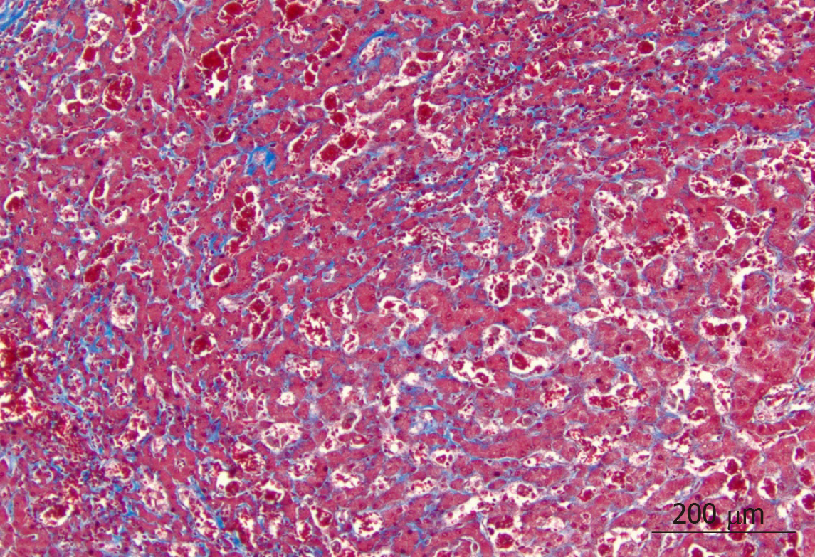
Figure 2 Trichrome stain highlighted extensive sinusoidal fibrosis and established portal-portal bridging fibrosis with evolution toward cirrhosis (× 10 magnification).
- Citation: Alkhayyat M, Saleh MA, Zmaili M, Sanghi V, Singh T, Rouphael C, Simons-Linares CR, Romero-Marrero C, Carey WD, Lindenmeyer CC. Successful liver transplantation for acute sickle cell intrahepatic cholestasis: A case report and review of the literature. World J Hepatol 2020; 12(3): 108-115
- URL: https://www.wjgnet.com/1948-5182/full/v12/i3/108.htm
- DOI: https://dx.doi.org/10.4254/wjh.v12.i3.108


