Copyright
©The Author(s) 2018.
World J Hepatol. Oct 27, 2018; 10(10): 780-784
Published online Oct 27, 2018. doi: 10.4254/wjh.v10.i10.780
Published online Oct 27, 2018. doi: 10.4254/wjh.v10.i10.780
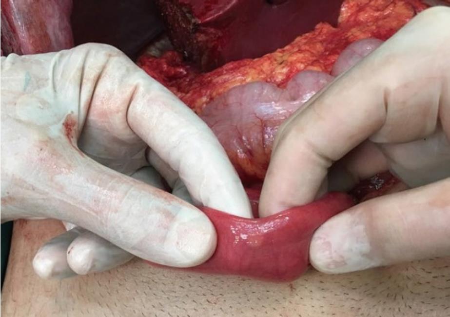
Figure 1 Intraoperative view of the tumor located in the antimesenteric border of the small intestine.
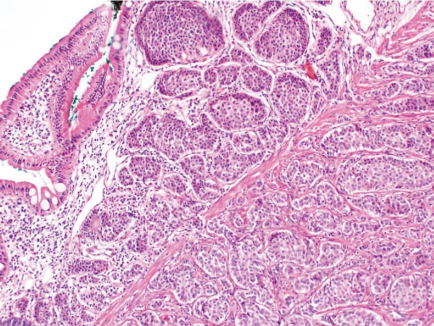
Figure 2 Tumor cells are seen in the submucosa with insulary pattern (HE × 100).
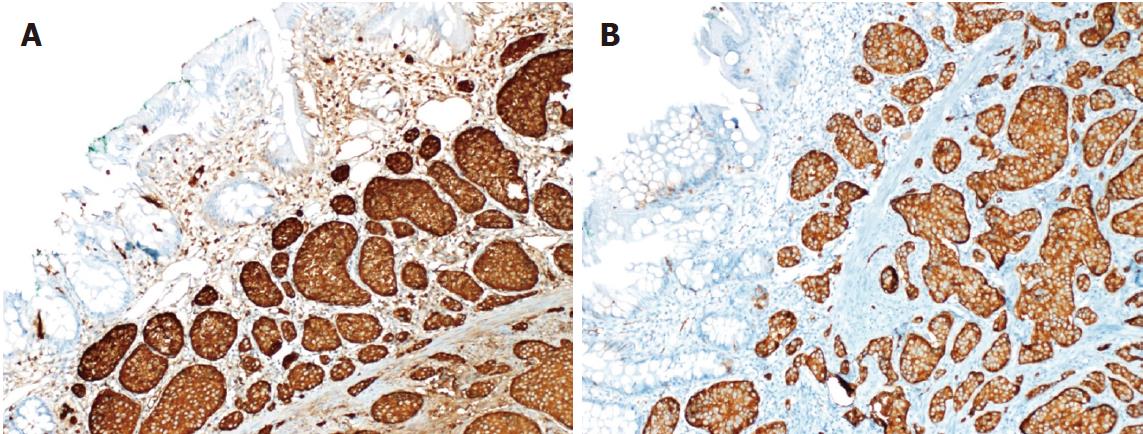
Figure 3 Positive immunostaining of tumor cells with chromogranin or synaptophysin antibody (HE × 100).
A: Chromogranin; B: Synaptophysin.
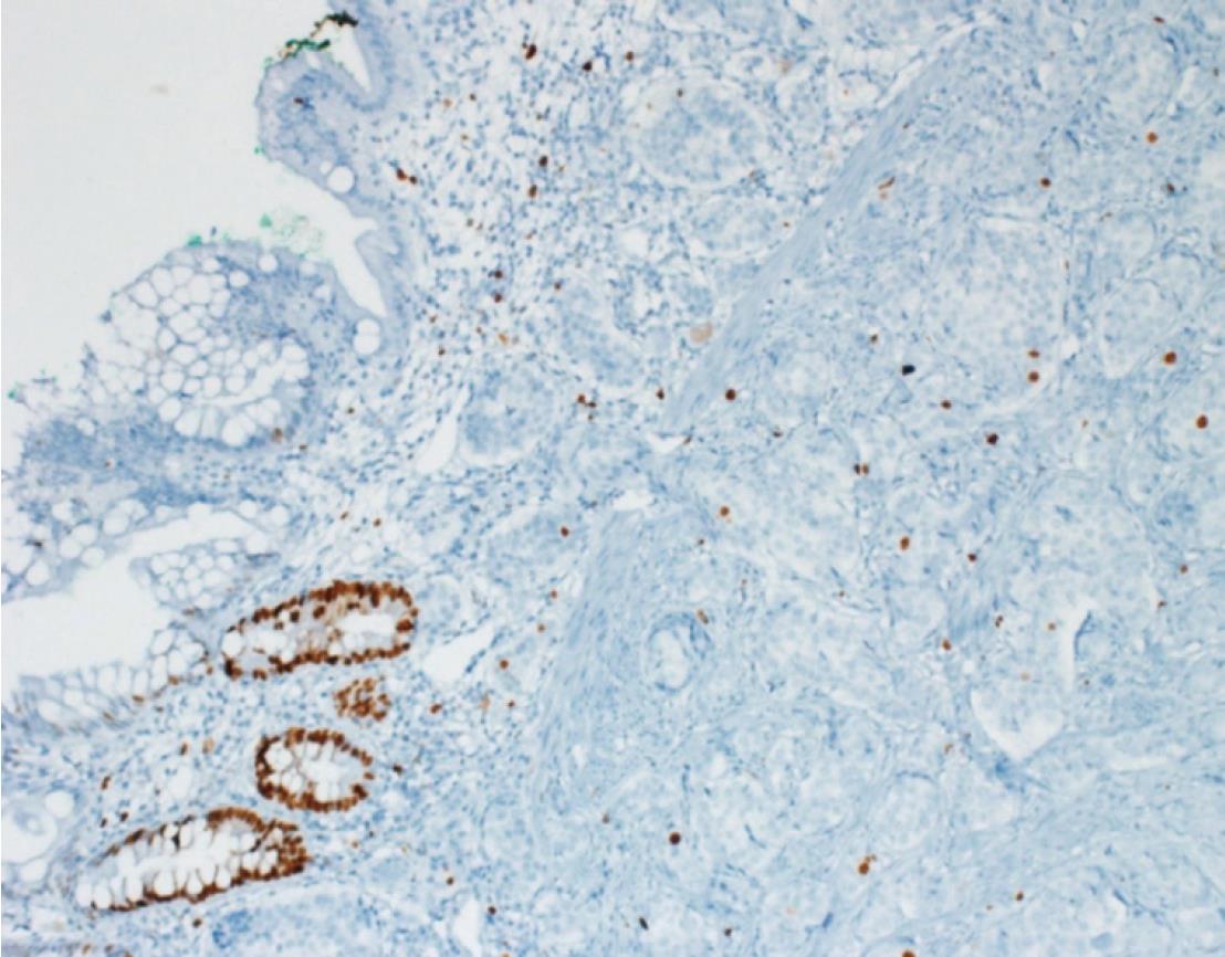
Figure 4 The low proliferation index in the tumor with Ki67 antibody (HE × 100).
- Citation: Akbulut S, Isik B, Cicek E, Samdanci E, Yilmaz S. Neuroendocrine tumor incidentally detected during living donor hepatectomy: A case report and review of literature. World J Hepatol 2018; 10(10): 780-784
- URL: https://www.wjgnet.com/1948-5182/full/v10/i10/780.htm
- DOI: https://dx.doi.org/10.4254/wjh.v10.i10.780


