Copyright
©The Author(s) 2021.
World J Stem Cells. Aug 26, 2021; 13(8): 1049-1057
Published online Aug 26, 2021. doi: 10.4252/wjsc.v13.i8.1049
Published online Aug 26, 2021. doi: 10.4252/wjsc.v13.i8.1049
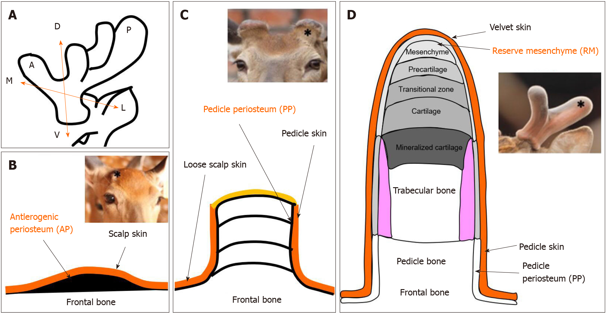
Figure 2 Schematic diagram of locations of antler stem cells.
A: Schematic diagram to show the three axes of the antler development: A ↔ P: Anterior-posterior axis; D ↔ V: Dorso-ventral axis; M ↔ L: Medio-lateral axis; B: The antlerogenic periosteum is present in the embryo and after birth as a localized thickening of the periosteum of the frontal bone; C: Regeneration of an antler initiated from the cells residing in the pedicle periosteum; D: Endochondral bone growth occurs at the distal tip, and cells in the reserve mesenchyme are responsible for rapid antler growth. Star in insert figure: Location of antler stem cells.
- Citation: Zhang W, Ke CH, Guo HH, Xiao L. Antler stem cells and their potential in wound healing and bone regeneration. World J Stem Cells 2021; 13(8): 1049-1057
- URL: https://www.wjgnet.com/1948-0210/full/v13/i8/1049.htm
- DOI: https://dx.doi.org/10.4252/wjsc.v13.i8.1049


