Copyright
©The Author(s) 2018.
World J Stem Cells. Nov 26, 2018; 10(11): 146-159
Published online Nov 26, 2018. doi: 10.4252/wjsc.v10.i11.146
Published online Nov 26, 2018. doi: 10.4252/wjsc.v10.i11.146
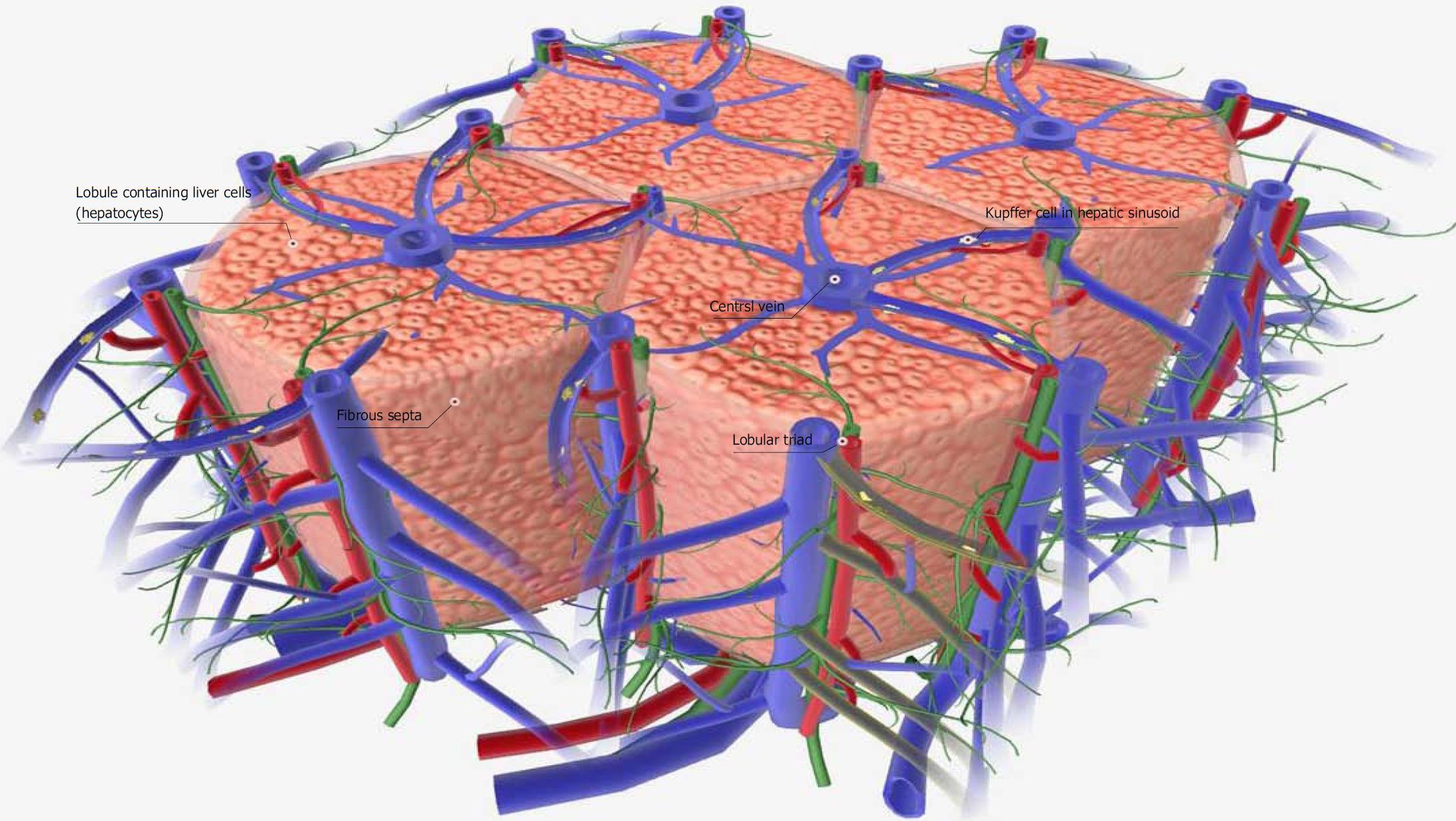
Figure 1 Schematic diagram of a normal liver tissue model.
The liver is a digestive organ that filters and detoxifies blood from the digestive tract. It also produces proteins, such as albumin, and synthesizes cholesterol and bile. The functional portion of the liver tissue is organized into hexagonal columns called liver lobules. Each liver lobule contains hundreds of individual liver cells (hepatocytes) and a large central vein. Lobular portal triads, which contain branches from the hepatic portal vein, hepatic artery and bile duct, are located at the points of the hexagonal lobule. Blood from the branches of the hepatic artery joins the blood of the hepatic vein branches, forming hepatic sinusoids. Hepatic sinusoids are lined with specialized cells called Kupffer cells, which help collect debris and detoxify the blood. All hepatic sinusoids in the liver lobule drain into the central vein. Adjacent lobules are separated by a thin fibrous septa. Images were obtained from BIODIGITAL HUMAN 3.0 (https://human.biodigital.com/index.html) (BioDigital, Broadway, NY, United States).
- Citation: Nahar S, Nakashima Y, Miyagi-Shiohira C, Kinjo T, Toyoda Z, Kobayashi N, Saitoh I, Watanabe M, Noguchi H, Fujita J. Cytokines in adipose-derived mesenchymal stem cells promote the healing of liver disease. World J Stem Cells 2018; 10(11): 146-159
- URL: https://www.wjgnet.com/1948-0210/full/v10/i11/146.htm
- DOI: https://dx.doi.org/10.4252/wjsc.v10.i11.146


