Copyright
©The Author(s) 2015.
World J Stem Cells. Aug 26, 2015; 7(7): 1047-1053
Published online Aug 26, 2015. doi: 10.4252/wjsc.v7.i7.1047
Published online Aug 26, 2015. doi: 10.4252/wjsc.v7.i7.1047
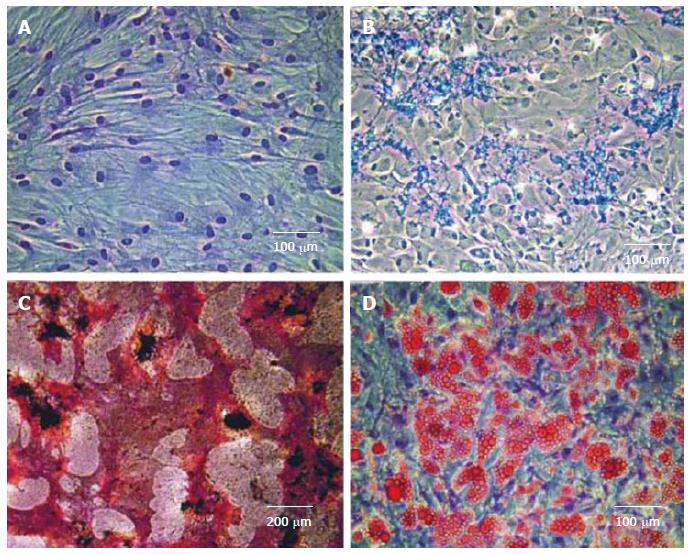
Figure 1 Mesenchymal stem cells are viewed as a yardstick of adult stem cells.
A: Human mesenchymal stem cells (MSCs) isolated from anonymous adult human bone marrow donor after culture expansion (H and E staining); B: Chondrocytes derived from human mesenchymal stem cells showing positive staining to Alcian blue. Additional molecular and genetic markers can be used to further characterize MSC-derived chondrocytes; C: Osteoblasts derived from human mesenchymal stem cells showing positive von Kossa staining for calcium deposition (black) and active alkaline phosphatase enzyme (red). Additional molecular and genetic markers can be used to further characterize MSC-derived chondrocytes; D: Adipocytes derived from human mesenchymal stem cells showing positive Oil Red-O staining of intracellular lipids. Additional molecular and genetic markers can be used to further characterize MSC-derived chondrocytes[16].
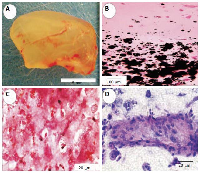
Figure 2 Engineered neogenesis of human-shaped mandibular condyle from mesenchymal stem cells.
A: Harvested osteochondral construct retained the shape and dimension of the cadaver human mandibular condyle after in vivo implantation; B: Von Kossa-stained section showing the interface between stratified chondral and osseous layers. Multiple mineralization nodules are present in the osseous layer (lower half of the photomicrograph), but absent in the chondral layer; C: Positive safranin O staining of the chondrogenic layer indicates the synthesis of abundant glycosaminoglycans; D: H and E-stained section of the osteogenic layer showing a representative osseous island-like structure consisting of MSC-differentiated osteoblast-like cells on the surface and in the center. Reproduced with permission from Biomedical Engineering Society[36].
Figure 3 Design and engineering of minipig mandibular condyle.
A: Original computed tomography scan of minipig mandible; B: Image-based design of condyle scaffold; C: Ppolycaprolactone degradable polymer scaffold fabricated with selective laser sintering attached to the ramus; D: Regrowth of condyle following 3 months' implantation (new condyle shown in red circle); E: Comparison with normal condyle from contralateral side in Yucatan minipig[37].
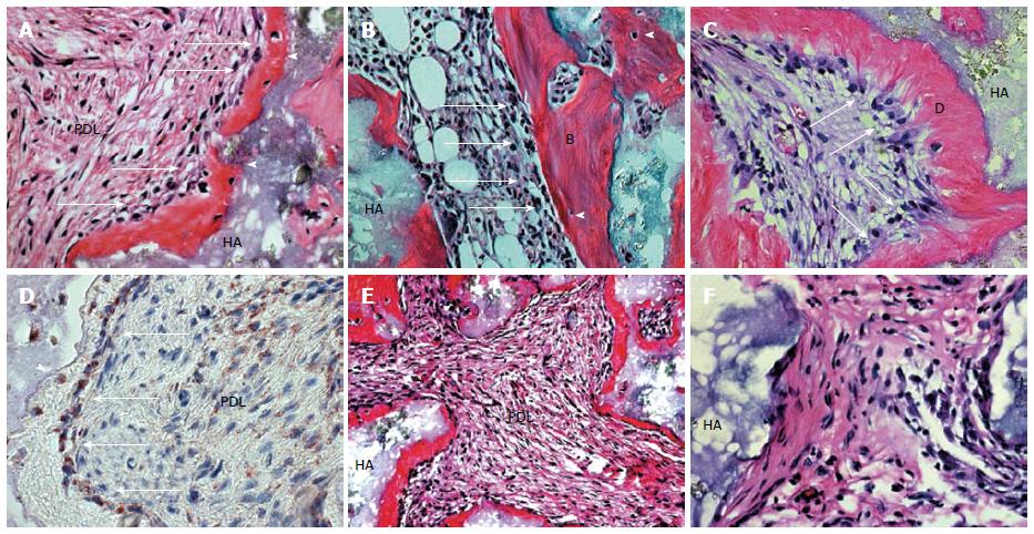
Figure 4 Generation of cementum-like and PDL-like structures in vivo by PDLSCs.
A: After 8 wk of transplantation, PDLSCs differentiated into cementoblast-like cells (arrows) that formed a cementum-like structure (C) on the surface of the hydroxyapatite tricalcium phosphate (HA) carrier; cementocyte-like cells (arrowhead) and PDL-like tissue (PDL) were also generated; B: BMSSC transplant was used as control to show the formation of a bone/marrow structure containing osteoblasts (arrows), osteocytes (arrowhead), and elements of bone (B) and haemopoietic marrow (HP); C: DPSC transplant was also used as a control to show a dentin/pulp-like structure containing odontoblasts (arrows) and dentin like (D) and pulp-like (Pulp) tissue; D: Immunohistochemical staining showed that PDLSCs generated cementum-like structure (C) and differentiated into cementoblast-like cells (arrows) and cementocyte-like cells (arrowhead) that stained positive for human-specific mitochondria antibody. Part of the PDL-like tissue (PDL) also stained positive for human specific mitochondria antibody (within dashed line); E: Of 13 selected strains of single-colony derived PDLSC, only eight (61%) generated cementum/PDL-like structures in vivo as shown at lower magnification (approximately 20). New cementum-like structure (C) formed adjacent to the surfaces of the carrier (HA) and associated with PDL-like tissue (PDL); F: The othser five strains did not generate mineralised or PDL-like tissues in vivo[38].
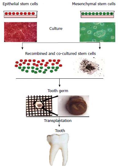
Figure 5 Use of stem cells for tooth formation in vitro and ex vivo.
A tooth germ can be created in vitro after co-culture of isolated epithelial and mesenchymal stem cells. This germ could be implanted into the alveolar bone and finally develop into a fully functional tooth[40].
- Citation: Aly LAA. Stem cells: Sources, and regenerative therapies in dental research and practice. World J Stem Cells 2015; 7(7): 1047-1053
- URL: https://www.wjgnet.com/1948-0210/full/v7/i7/1047.htm
- DOI: https://dx.doi.org/10.4252/wjsc.v7.i7.1047


